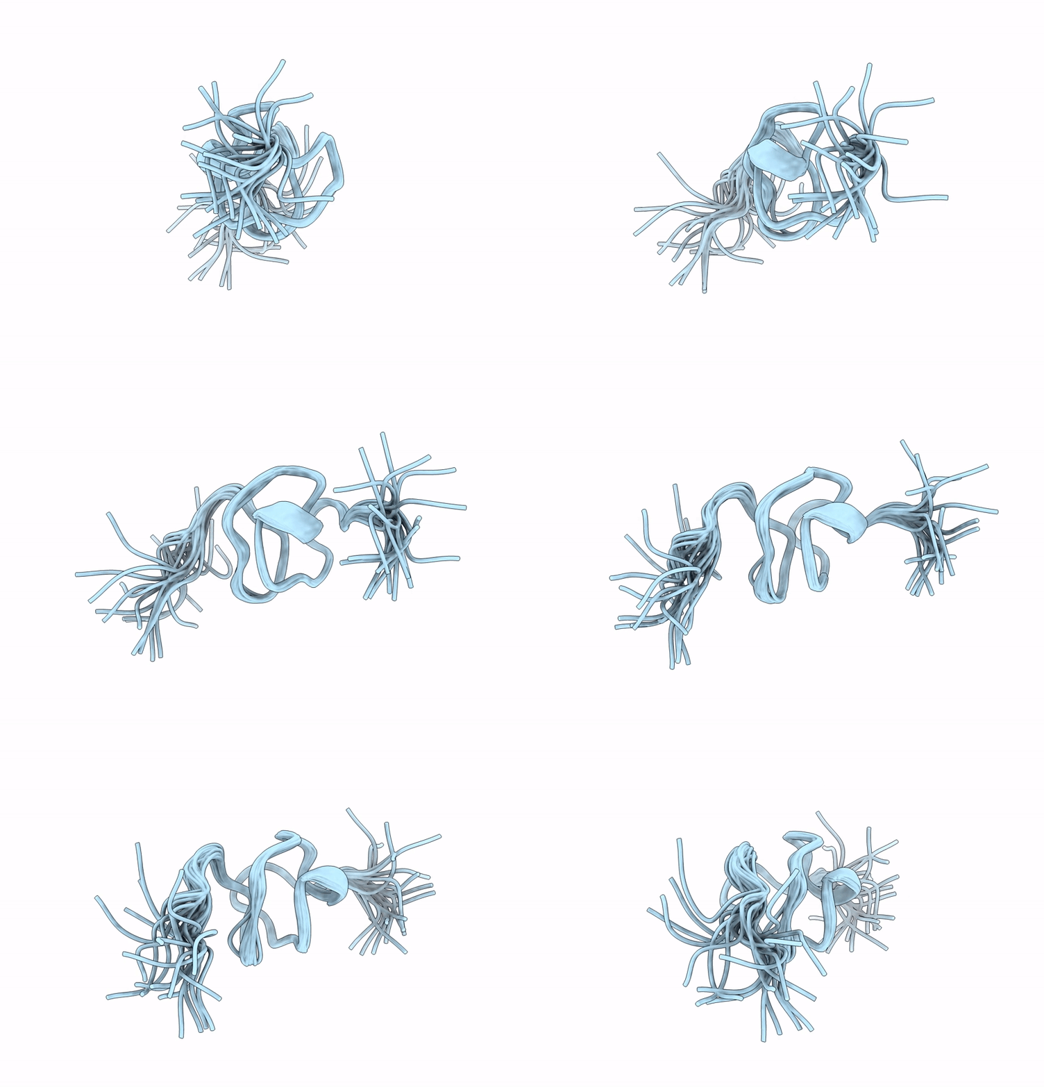
9NVH image
Entry Detail
PDB ID:
9NVH
Keywords:
Title:
NMR structure of conotoxin k-SrXIA
Biological Source:
Source Organism:
PDB Version:
Deposition Date:
2025-03-20
Release Date:
2025-05-14
Method Details:
Experimental Method:
Conformers Calculated:
200
Conformers Submitted:
20
Selection Criteria:
structures with the lowest energy
Macromolecular Entities
Polymer Type:polypeptide(L)
Description:Kappa-conotoxin SrXIA
Chain IDs:A
Chain Length:35
Number of Molecules:1
Biological Source:Conus spurius
Ligand Molecules
Primary Citation
Block of Kv1 potassium channels and NMR structure of recombinant conotoxin kappa-SrXIA.
Toxicon 262 108384 108384 (2025)
PMID: 40324600 DOI: 10.1016/j.toxicon.2025.108384

Abstact

The biologically active components of Conus venoms are mainly small peptides with disulfide-bonded structures. Some conotoxins contain post-translational modifications as an evolutionary strategy to enhance their potency and selectivity towards ion channels and receptors. Few conotoxins are known to target mammalian Kv1 channels. κ-SrXIA from the venom of Conus spurius inhibits the voltage-gated potassium Kv1.2 and Kv1.6 channels through a basic ring of Arg. The 32 amino acid κ-SrXIA has eight Cys residues arranged in the pattern that defines the I-superfamily with four disulfide bridges, two gamma-carboxy-glutamates (Gla), and a Pro-amidated C-terminus (X). In this study, we obtained and determined the biological activity and NMR structure of recombinant κ-SrXIA without Gla and X. The conotoxin cDNA was expressed in E. coli CD41 and purified by GST-affinity chromatography and RP-HPLC. Pharmacological assays were performed by two-electrode voltage-clamp recordings in Xenopus laevis oocytes expressing recombinant Kv1.1, Kv1.2, Kv1.3, Kv1.4 and Kv1.6 channels. Except for Kv1.3, κ-Sr-XIA irreversibly blocked Kv1 channels displaying a lower affinity and a slower inhibition kinetics than native conotoxin. Even when κ-SrXIA displayed a high structural similarity to GXIA from the I3-superfamily, the resultant disulfide connectivity forming an ICK+1 motif showed two antiparallel β-strands as ι-RXIA from the I1-superfamily. This study represents the first 3D NMR structure for a member of the I2-superfamily.

Legend

Protein

Chemical

Disease

Primary Citation of related structures