Deposition Date
2025-03-13
Release Date
2025-08-27
Last Version Date
2025-11-12
Entry Detail
PDB ID:
9NR0
Keywords:
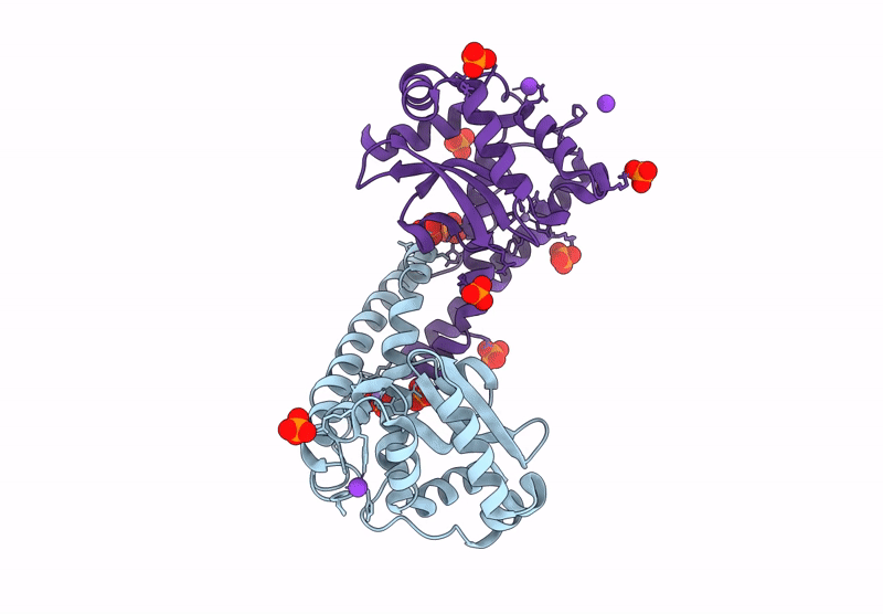
Title:
Finding the exit route of hydrogen peroxide from the manganese superoxide dismutase (MnSOD) active site
Biological Source:
Source Organism:
Homo sapiens (Taxon ID: 9606)
Host Organism:
Method Details:
Experimental Method:
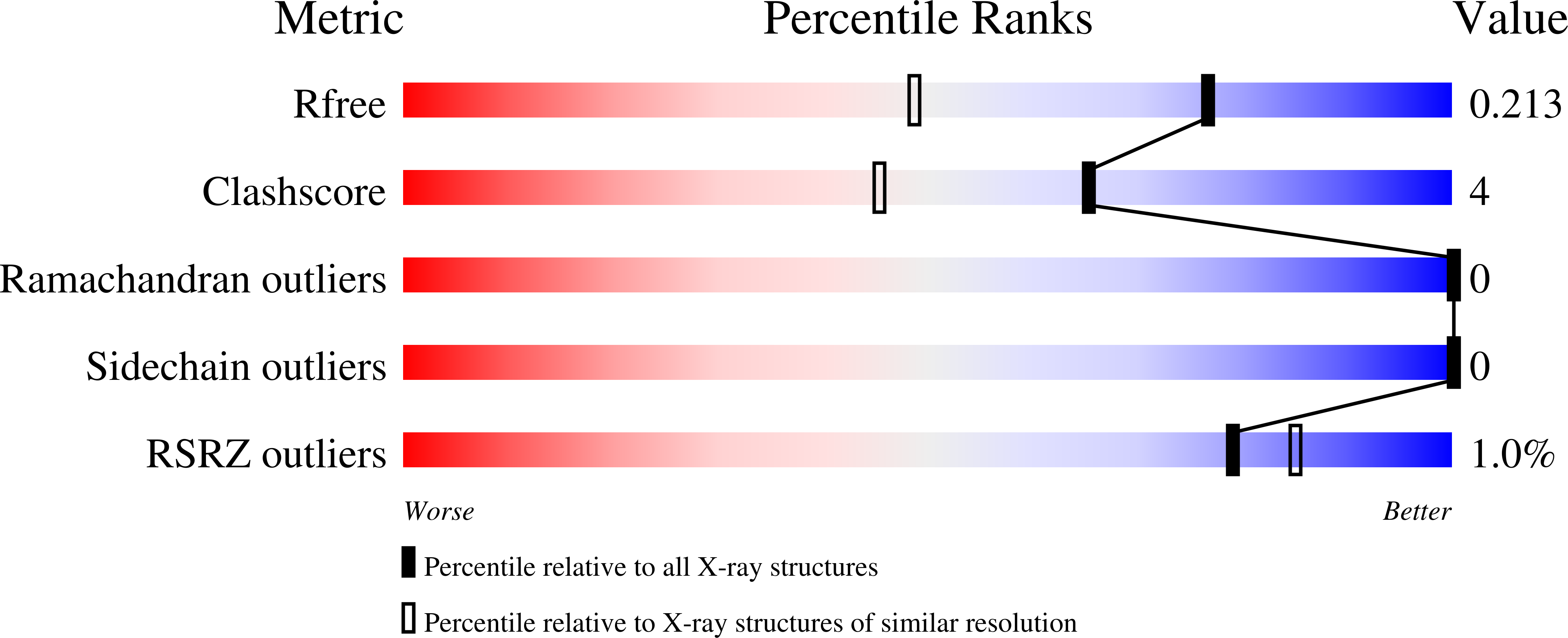
Resolution:
1.55 Å
R-Value Free:
0.21
R-Value Work:
0.17
R-Value Observed:
0.17
Space Group:
P 61 2 2