Deposition Date
2025-03-11
Release Date
2025-08-27
Last Version Date
2025-08-27
Entry Detail
PDB ID:
9NPG
Keywords:
Title:
X-ray crystal structure of recombinant Can f 1-C100S in complex with human IgE mAb 12F3 Fab
Biological Source:
Source Organism:
Homo sapiens (Taxon ID: 9606)
Canis lupus familiaris (Taxon ID: 9615)
Canis lupus familiaris (Taxon ID: 9615)
Host Organism:
Method Details:
Experimental Method:
Resolution:
3.12 Å
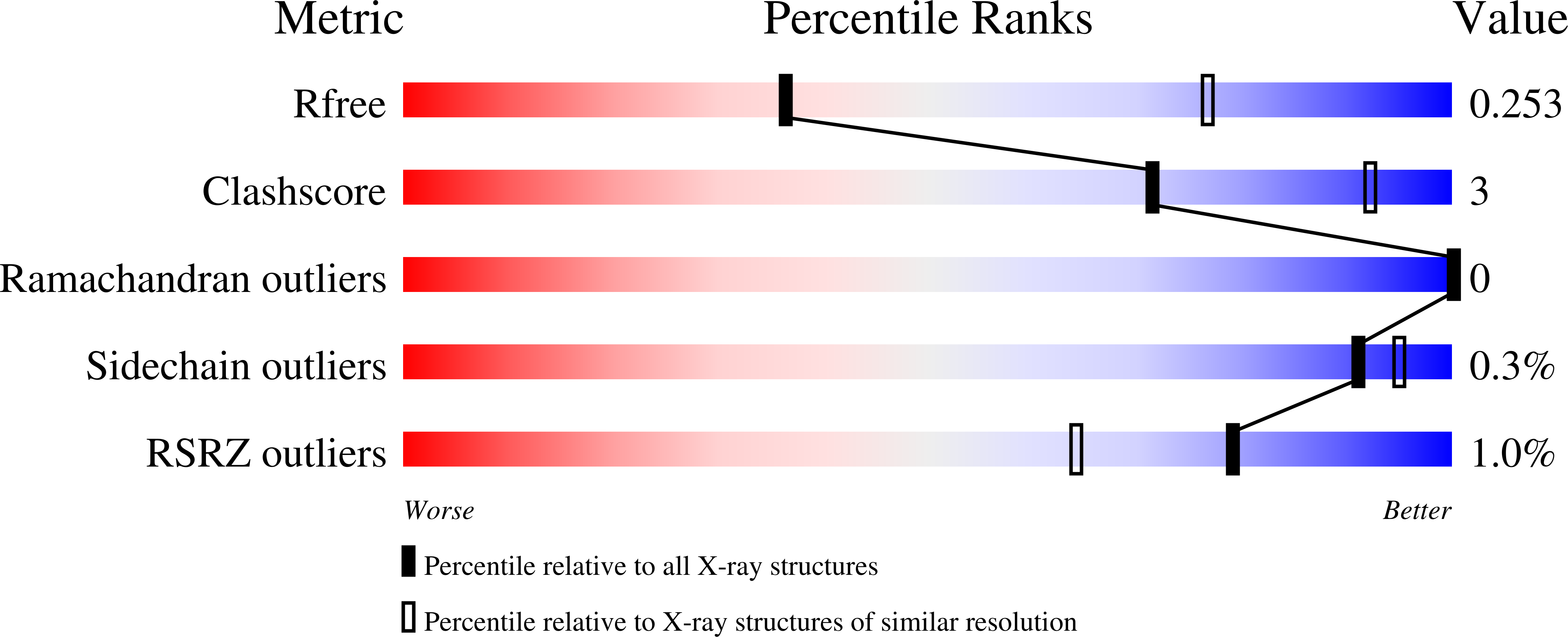
R-Value Free:
0.24
R-Value Work:
0.20
R-Value Observed:
0.20
Space Group:
P 1 21 1