9N2K image
Deposition Date 2025-01-29
Release Date 2026-01-07
Last Version Date 2026-01-14
Entry Detail
PDB ID:
9N2K
Title:
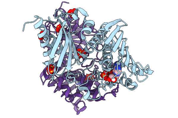
N-terminal domain of Bacillus subtilis MutL bound to ADP
Biological Source:
Source Organism(s):
Expression System(s):
Method Details:
Experimental Method:
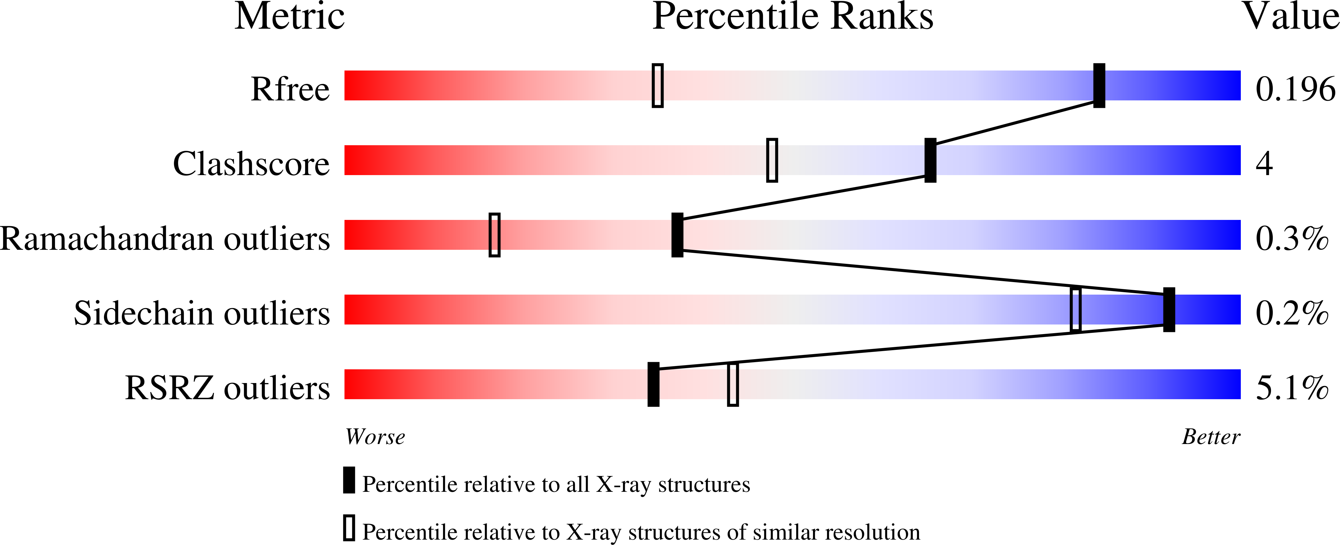
Resolution:
1.36 Å
R-Value Free:
0.19
R-Value Work:
0.17
R-Value Observed:
0.17
Space Group:
P 1 21 1
Macromolecular Entities
Structures with similar UniProt ID
Protein Blast
Polymer Type:polypeptide(L)
Molecule:DNA mismatch repair protein MutL
Gene (Uniprot):mutL
Chain IDs:A, B
Chain Length:335
Number of Molecules:2
Biological Source:Bacillus subtilis
Primary Citation
Bacillus subtilis MutL samples multiple conformations during nucleotide binding and hydrolysis.
Structure ? ? ? (2025)
PMID: 41478286 DOI: 10.1016/j.str.2025.12.007

Abstact

DNA mismatch repair is an evolutionarily conserved repair pathway that corrects replication errors, thereby preventing genome instability. Two evolutionarily conserved proteins, MutS and MutL, recognize the mismatch and mark the newly synthesized strand for repair. Previous studies have shown how bacterial MutS homodimers function asymmetrically to recognize mismatches and recruit MutL. However, whether MutL homodimers also function asymmetrically to coordinate binding to MutS and activation of their nuclease activity remains unclear. Here, we characterize the ATPase domain of Bacillus subtilis MutL, a MutL protein with endonuclease activity, and delineate the differences with Escherichia coli MutL, a homolog without endonuclease activity. We find that B. subtilis MutL has low affinity for ATP and samples a repertoire of conformations that resemble those observed in eukaryotic MutL paralogs, indicating a relationship between ATP-induced dimer compaction and nuclease activity.

Legend

Protein

Chemical

Disease

Primary Citation of related structures
Feedback Form
Name
Email
Institute
Feedback