9MEX image
Deposition Date 2024-12-09
Release Date 2025-05-14
Last Version Date 2025-06-25
Entry Detail
PDB ID:
9MEX
Keywords:
Title:
Structure of phosphocysteine intermediate of human PRL1 phosphatase
Biological Source:
Source Organism:
Homo sapiens (Taxon ID: 9606)
Method Details:
Experimental Method:
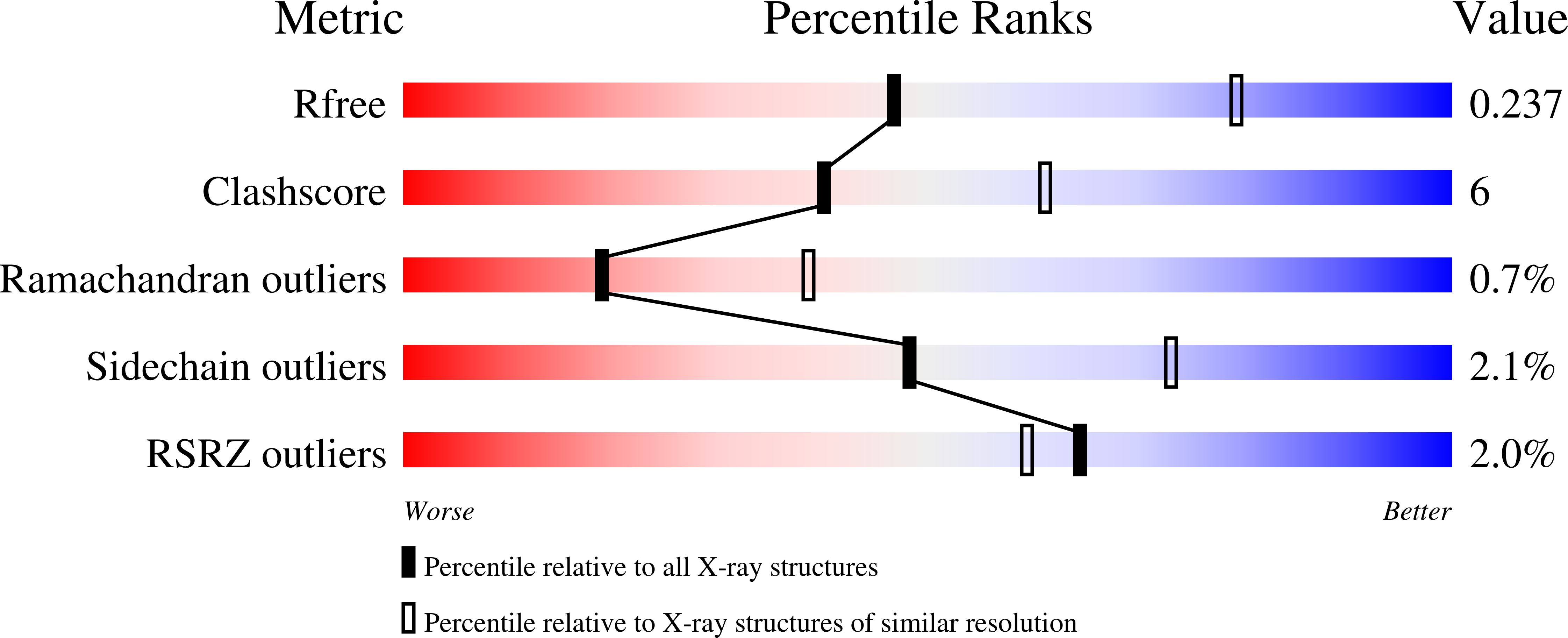
Resolution:
2.60 Å
R-Value Free:
0.23
R-Value Work:
0.19
R-Value Observed:
0.19
Space Group:
I 21 3
Macromolecular Entities
Polymer Type:polypeptide(L)
Molecule:Protein tyrosine phosphatase type IVA 1
Gene (Uniprot):PTP4A1
Mutations:C49S, D72A
Chain IDs:A, B
Chain Length:164
Number of Molecules:2
Biological Source:Homo sapiens
Modified Residue
Compound ID Chain ID Parent Comp ID Details 2D Image
CSP A CYS modified residue
Ligand Molecules
Primary Citation
Structure of the phosphocysteine intermediate of the phosphatase of regenerating liver PTP4A1.
J.Biol.Chem. 301 110251 110251 (2025)
PMID: 40398601 DOI: 10.1016/j.jbc.2025.110251

Abstact

Phosphatases of regenerating liver (PRL or PTP4A) are protein phosphatases implicated in cell growth, magnesium homeostasis, and cancer metastasis. During catalysis, a phosphocysteine intermediate forms, which must undergo hydrolysis to regenerate the active enzyme. In addition to dephosphorylating substrates, PRLs act as pseudo-phosphatases and bind CBS-pair domain divalent metal cation transport mediators (CNNMs) to regulate magnesium transport. In this study, we investigate the role of PRL residues in phosphocysteine hydrolysis using mutagenesis, enzyme assays, and X-ray crystallography. Loss of an aspartic acid and cysteine in the catalytic site disrupts hydrolysis and stabilizes the phosphocysteine intermediate for weeks. We use this C49S/D72A double mutant to determine the crystal structure of the cysteine phosphorylated form of PRL1 (PTP4A1). The structure confirms that phosphocysteine sterically interferes with CNNM binding, consistent with previous biochemical studies. In vitro enzyme assays reveal the aspartic acid mutation increases the initial rate of catalysis for all three PRL paralogs while the homologous mutation in the phosphatases, PTP1B and PTPN12, disrupts catalysis. This highlights the mechanistic differences between PRLs and classical protein tyrosine phosphatases. Our findings refine our understanding of PRL catalysis and identify novel mutations for investigating PRL function in cancer and magnesium homeostasis.

Legend

Protein

Chemical

Disease

Primary Citation of related structures