Deposition Date
2025-03-17
Release Date
2025-07-30
Last Version Date
2025-07-30
Entry Detail
PDB ID:
9MBL
Keywords:
Title:
2-Oxo-dATP hydrolysis in human MTH1(G2K mutant) crystal using Mn2+: the ES-2M complex
Biological Source:
Source Organism(s):
Homo sapiens (Taxon ID: 9606)
Expression System(s):
Method Details:
Experimental Method:
Resolution:
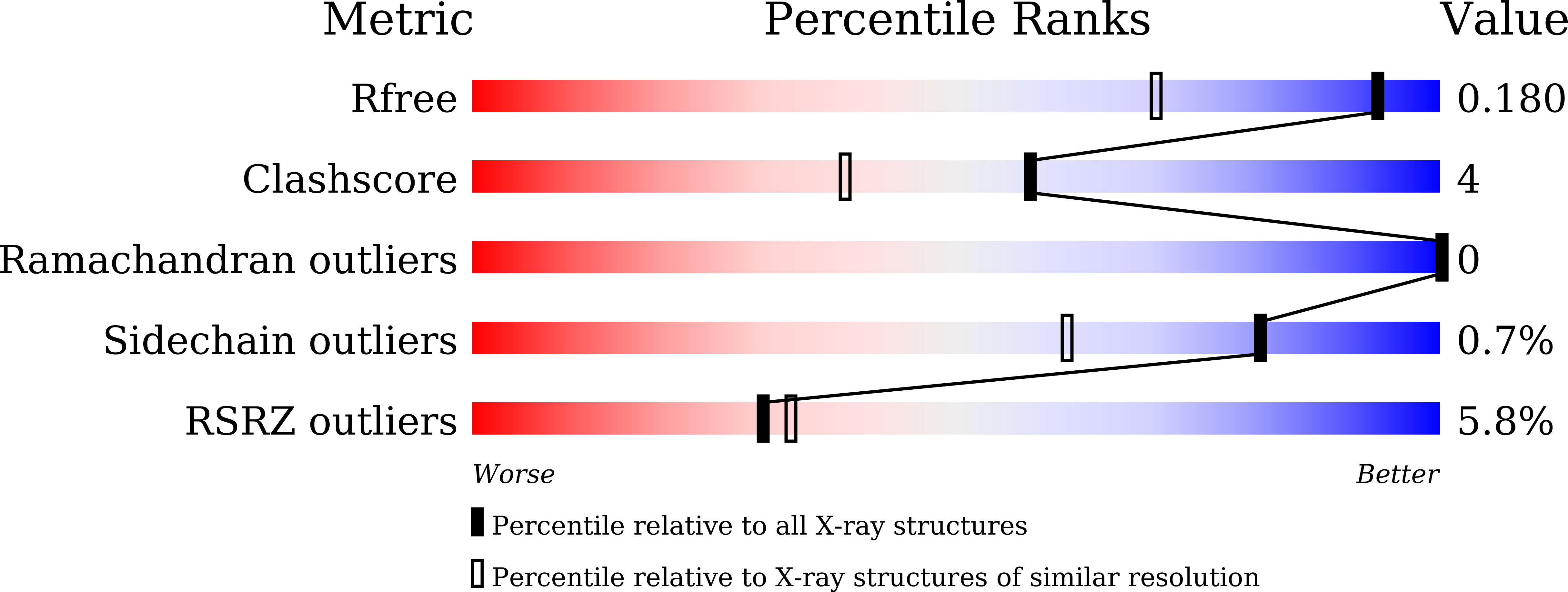
1.37 Å
R-Value Free:
0.18
R-Value Work:
0.13
R-Value Observed:
0.13
Space Group:
P 21 21 21