9M7L image
Deposition Date 2025-03-10
Release Date 2025-06-18
Last Version Date 2025-07-02
Entry Detail
PDB ID:
9M7L
Title:
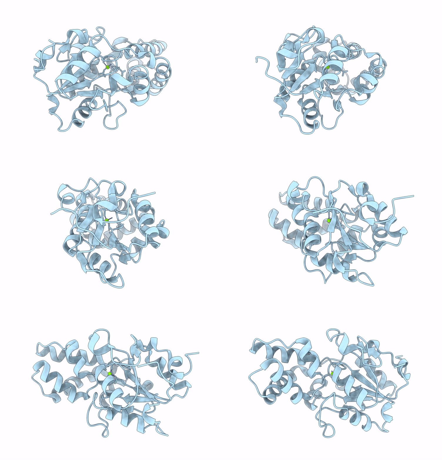
Crystal structure of Pseudouridine 5'-monophosphate phosphatase from human (hHDHD1A) in the unliganded state
Biological Source:
Source Organism:
Homo sapiens (Taxon ID: 9606)
Method Details:
Experimental Method:
Resolution:
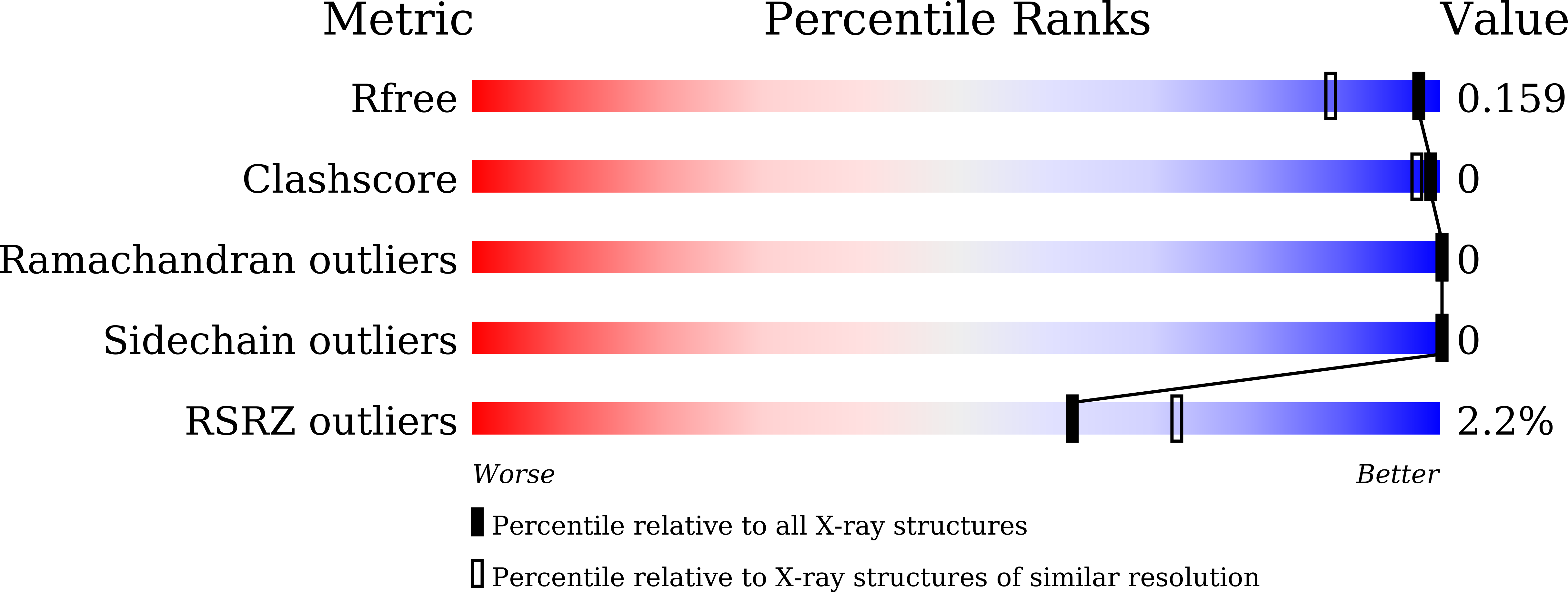
1.36 Å
R-Value Free:
0.15
R-Value Work:
0.14
R-Value Observed:
0.14
Space Group:
P 2 21 21
Macromolecular Entities
Polymer Type:polypeptide(L)
Molecule:Pseudouridine-5'-phosphatase
Gene (Uniprot):PUDP
Chain IDs:A
Chain Length:226
Number of Molecules:1
Biological Source:Homo sapiens
Ligand Molecules
Primary Citation
Structural and functional insights into the substrate specificity of the pseudouridine monophosphate phosphatase HDHD1A.
J.Biol.Chem. 301 110257 110257 (2025)
PMID: 40409548 DOI: 10.1016/j.jbc.2025.110257

Abstact

Pseudouridine (Ψ) is one of the most abundant RNA modifications. Following RNA degradation, Ψ nucleotides are dephosphorylated and catabolized into uracil and ribose 5'-phosphate via a two-step enzymatic reaction catalyzed by enzymes present in many bacteria and eukaryotes, but not in mammals. Malfunction of Ψ catabolism has adverse physiological effects in plants. In humans, the enzyme HDHD1A dephosphorylates pseudouridine 5'-monophosphate (ΨMP), and the resulting Ψ is excreted in the urine. In this study, we determined the crystal structures of human HDHD1A (hHDHD1A) complexed with Ψ. The structure of hHDHD1A consists of a catalytic domain with a Rossmann α/β-fold and a cap domain, with a magnesium ion at the junction of the two domains. Ψ is bound to the active site in an orientation where its nucleobase, uracil-Ψ, is surrounded by the cap domain residues, and the ribose moiety is located next to the Mg2+-binding site. The active site is composed mainly of hydrophobic residues but two essential charged residues, Glu23 and Lys46, are present in the vicinity of uracil-Ψ. Glu23 interacts with the Ψ-specific N1 atom, while Lys46 interacts with the O2 atom of uracil-Ψ. Mutagenesis and kinetic analysis indicated that active site residues are involved in substrate binding and/or catalysis. In addition to Ψ-specific hydrophilic interactions, shape complementarity between ΨMP and the active site pocket is a key element underlying substrate specificity in hHDHD1A. This study provided structural and functional insights into the substrate specificity of hHDHD1A for ΨMP, highlighting both similarities and differences compared to other Ψ-recognizing enzymes.

Legend

Protein

Chemical

Disease

Primary Citation of related structures