9M1A image
Deposition Date 2025-02-25
Release Date 2025-06-11
Last Version Date 2025-06-25
Entry Detail
PDB ID:
9M1A
Keywords:
Title:
Vitamin D receptor complex with a diethyldiphenylsilane derivative
Biological Source:
Source Organism:
Rattus norvegicus (Taxon ID: 10116)
Homo sapiens (Taxon ID: 9606)
Host Organism:
Method Details:
Experimental Method:
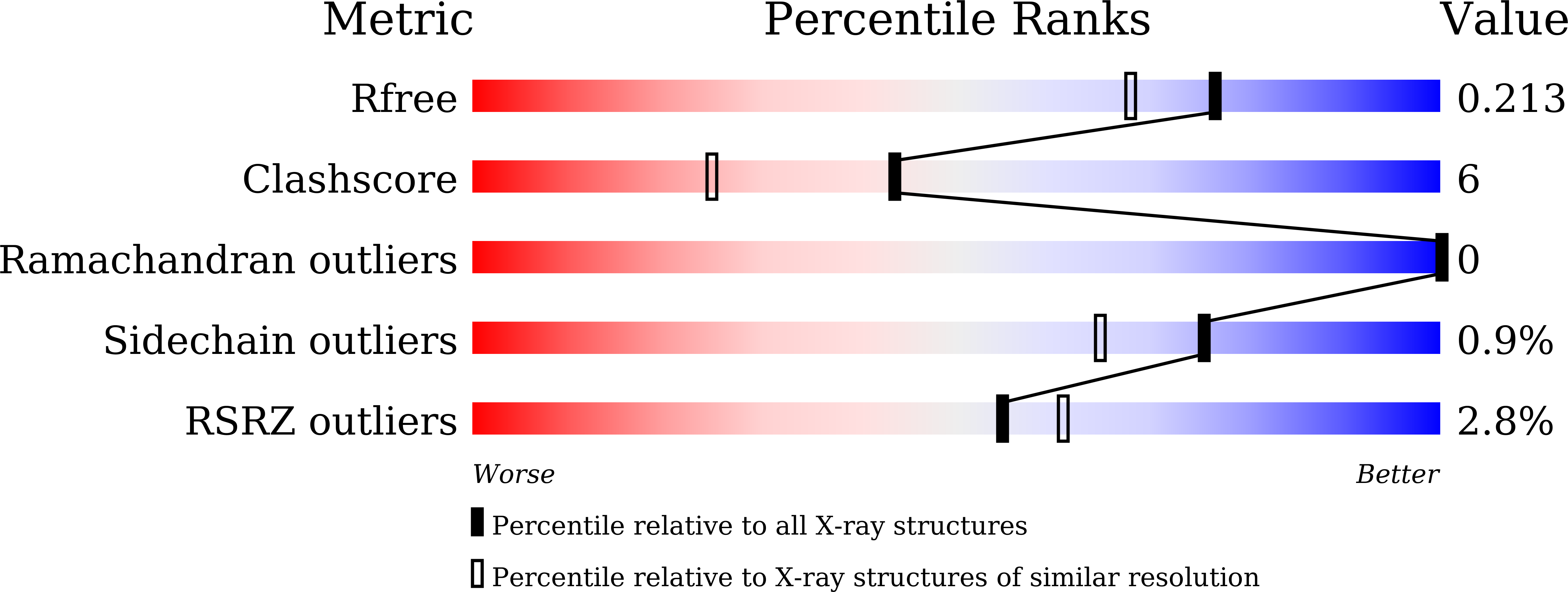
Resolution:
1.78 Å
R-Value Free:
0.21
R-Value Work:
0.17
R-Value Observed:
0.18
Space Group:
C 1 2 1
Macromolecular Entities
Structures with similar UniProt ID
Protein Blast
Polymer Type:polypeptide(L)
Molecule:Vitamin D3 receptor
Gene (Uniprot):Vdr
Chain IDs:A
Chain Length:271
Number of Molecules:1
Biological Source:Rattus norvegicus
Structures with similar UniProt ID
Protein Blast
Polymer Type:polypeptide(L)
Molecule:Mediator of RNA polymerase II transcription subunit 1
Gene (Uniprot):MED1
Chain IDs:B (auth: C)
Chain Length:13
Number of Molecules:1
Biological Source:Homo sapiens
Ligand Molecules
Primary Citation
Structure-activity relationship and crystallographic analyses of non-secosteroidal vitamin D receptor ligands bearing diphenylsilane core as a hydrophobic pharmacophore.
Bioorg.Med.Chem. 128 118261 118261 (2025)
PMID: 40494220 DOI: 10.1016/j.bmc.2025.118261

Abstact

Vitamin D receptor (VDR) is an attractive target of drug discovery for multiple diseases. In this study, we systematically designed and synthesized a series of diphenylsilane derivatives with diverse hydrophobic substituents and investigated their structure-activity relationship (SAR) as VDR agonists. The SAR study revealed that the activity is dependent on the type of substituent and the position of substitution, and the diethyl-di-m-tolylsilane scaffold was identified as the most suitable hydrophobic core structure of this type of VDR ligands. Interestingly, the small structural difference between n-propyl and allyl substituents resulted in a large difference in the activity. Comparison of the co-crystal structures of 14 diphenylsilane compounds, including less potent compounds, bound to the rat VDR ligand-binding domain suggested that the differences in activity are due to a combination of factors, including differences in hydrophilic and hydrophobic interactions, and ligand conformations.

Legend

Protein

Chemical

Disease

Primary Citation of related structures