9LYO image
Deposition Date 2025-02-20
Release Date 2025-04-02
Last Version Date 2025-04-02
Entry Detail
PDB ID:
9LYO
Keywords:
Title:
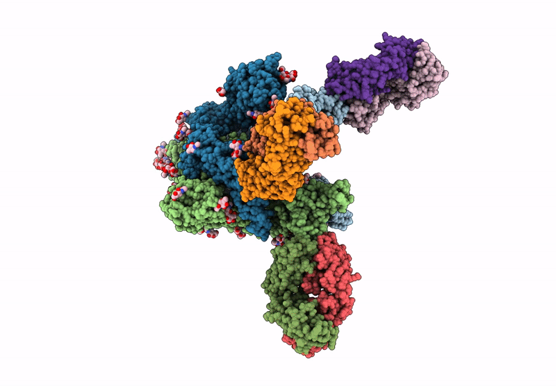
Alpha SARS-CoV-2 spike protein in complex with REGN10987 Fab homologue.
Biological Source:
Host Organism:
Method Details:
Experimental Method:
Resolution:
3.07 Å
Aggregation State:
PARTICLE
Reconstruction Method:
SINGLE PARTICLE
Macromolecular Entities
Polymer Type:polypeptide(L)
Molecule:Spike glycoprotein
Gene (Uniprot):S
Chain IDs:C (auth: A), D (auth: B), E (auth: C)
Chain Length:1286
Number of Molecules:3
Biological Source:Severe acute respiratory syndrome coronavirus 2
Polymer Type:polypeptide(L)
Molecule:REGN10987 Fab homologue (Light chain)
Chain IDs:A (auth: i), F (auth: g), H (auth: l)
Chain Length:218
Number of Molecules:3
Biological Source:Homo sapiens
Polymer Type:polypeptide(L)
Molecule:REGN10987 Fab homologue (Heavy chain)
Chain IDs:B (auth: j), G (auth: h), I (auth: n)
Chain Length:223
Number of Molecules:3
Biological Source:Homo sapiens
Primary Citation
Structure and dynamics of Alpha B.1.1.7 SARS-CoV-2 S-protein in complex with Fab of neutralizing antibody REGN10987.
Biochem.Biophys.Res.Commun. 755 151558 151558 (2025)
PMID: 40043614 DOI: 10.1016/j.bbrc.2025.151558

Abstact

One of the approaches for treatment of COVID-19 is a use of neutralizing antibodies (nAbs). The study of the mechanisms by which nAbs recognize different strains of SARS-CoV-2 may facilitate the development of new drugs and vaccines against the coronavirus infection. In this work, we present the 3.1 Å resolution cryo-electron microscopy structure of a full-length trimeric spike-protein (S-protein) of the SARS-CoV-2 Alpha (B.1.1.7) variant in complex with the Fab of the REGN10987 nAb. In the complex, two receptor-binding domains (RBDs) of the S-protein were observed in the 'up' state, whereas third RBD was in the 'down' state. This distinguishes the obtained structure from the complex of Delta (B.1.617.2) S-protein with REGN10987-Fab, where only one RBD was in the 'up' state. Probably some of the substituted residues (K478T, A570D, and S982A) located at the interprotomer interfaces are responsible for the greater Alpha S-protein opening upon the REGN10987-Fab binding. The Fab identically binds to the RBD in the both 'up' and 'down' conformations. The RBD-Fab interaction interface was refined to a resolution of 3.6 Å. The antibody binds to the receptor-binding motif (RBM), which prevents the S-protein from the binding to its receptor, angiotensin-converting enzyme 2 (ACE-2). Comparison with the structures of the Wuhan (wild type) and Delta RBD variants in complex with REGN10987-Fab revealed that the N501Y and T478K/L452R mutations presented in the RBM of the Alpha and Delta variants, respectively, do not affect the mode of the RBD-Fab interaction.

Legend

Protein

Chemical

Disease

Primary Citation of related structures