
Deposition Date
2025-02-17
Release Date
2025-07-02
Last Version Date
2025-09-10
Entry Detail
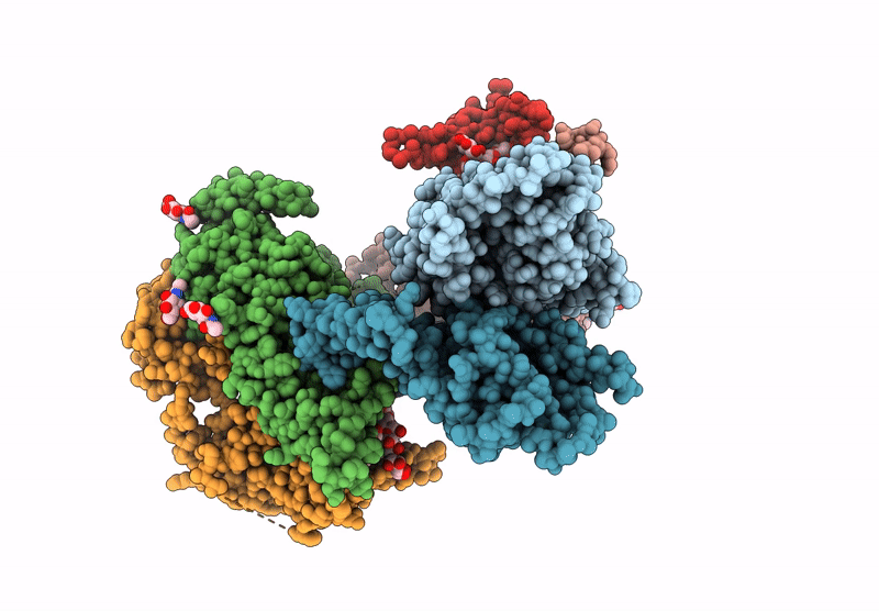
PDB ID:
9LWU
Keywords:
Title:
Cryo-EM structure of HRD1-SEL1LX3-XTP3B complex in C2 symmetry
Biological Source:
Source Organism(s):
Homo sapiens (Taxon ID: 9606)
Expression System(s):
Method Details:
Experimental Method:
Resolution:
3.50 Å
Aggregation State:
PARTICLE
Reconstruction Method:
SINGLE PARTICLE
