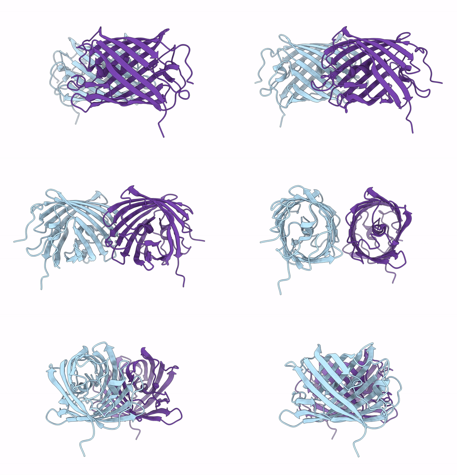
9LPU image
Deposition Date 2025-01-26
Release Date 2025-04-23
Last Version Date 2025-05-14
Entry Detail
PDB ID:
9LPU
Title:
Crystal structure of tKeima at pH 4.0
Biological Source:
Source Organism:
Method Details:
Experimental Method:
Resolution:
2.20 Å
R-Value Free:
0.27
R-Value Work:
0.23
R-Value Observed:
0.23
Space Group:
P 21 21 2
Macromolecular Entities
Polymer Type:polypeptide(L)
Molecule:Large stokes shift fluorescent protein
Gene (Uniprot):t-keima
Chain IDs:A, B
Chain Length:224
Number of Molecules:2
Biological Source:Montipora sp. 20
Modified Residue
Compound ID Chain ID Parent Comp ID Details 2D Image
CRQ A GLN chromophore
Primary Citation
pH-Induced Conformational Change of the Chromophore of the Large Stokes Shift Fluorescent Protein tKeima.
Molecules 30 ? ? (2025)
PMID: 40286219 DOI: 10.3390/molecules30071623

Abstact

Fluorescent proteins (FPs) are widely used as optical probes in molecular and cell biology. tKeima is a tetrameric, large Stokes shift red fluorescent protein and the ancestral protein of mt-Keima, which is widely applied as a pH-sensitive fluorescent probe. While the pH sensitivity of mt-Keima is well characterized, the pH-dependent properties of the ancestral tKeima have not been comprehensively elucidated. To obtain a better understanding of the effects of pH on tKeima, its fluorescent emission intensity at various pH levels was measured, and its crystal structure at pH 4.0 was determined at a resolution of 2.2 Å. The fluorescence emission intensity of tKeima at pH 4.0 decreased by approximately 65% compared with its peak emission at pH 10.0. The crystal structure of tKeima at pH 4.0 revealed both cis and trans conformations of the chromophore, in contrast to previously determined structures at pH 8.0, which showed only the cis conformation. This indicates that pH induces a conformational change of the chromophore in tKeima. Both the cis and trans conformations in tKeima were stabilized by hydrogen bonds with neighboring residues. A comparison of tKeima at pH 4.0 with tKeima at basic pH, as well as with mKeima, highlights its unique structural properties. These results provide a deeper understanding of the structural basis for the pH-induced fluorescence emission changes in the Keima family.

Legend

Protein

Chemical

Disease

Primary Citation of related structures