Deposition Date
2025-01-21
Release Date
2025-12-10
Last Version Date
2025-12-10
Entry Detail
PDB ID:
9LNJ
Keywords:
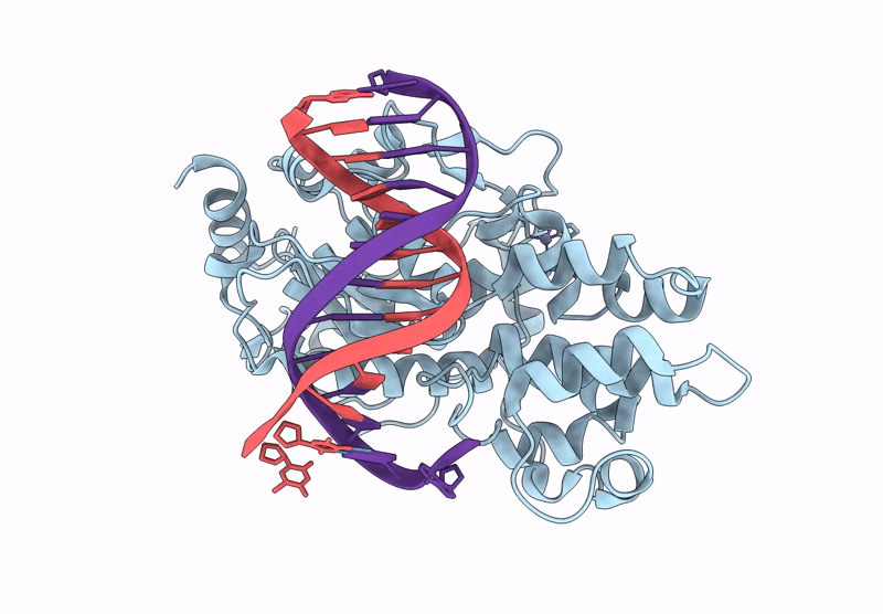
Title:
Crystal structure of VANC21 in complex with its target DNA (5-bromouridine substituted).
Biological Source:
Source Organism(s):
Arabidopsis thaliana (Taxon ID: 3702)
Expression System(s):
Method Details:
Experimental Method:
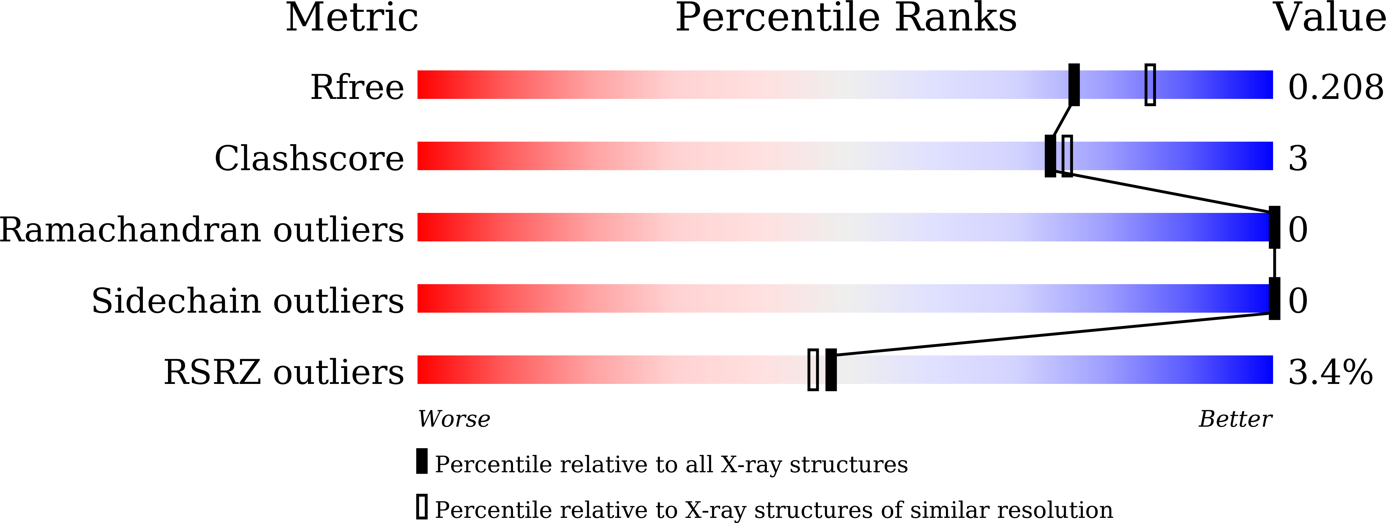
Resolution:
2.00 Å
R-Value Free:
0.20
R-Value Work:
0.17
R-Value Observed:
0.17
Space Group:
P 1 21 1