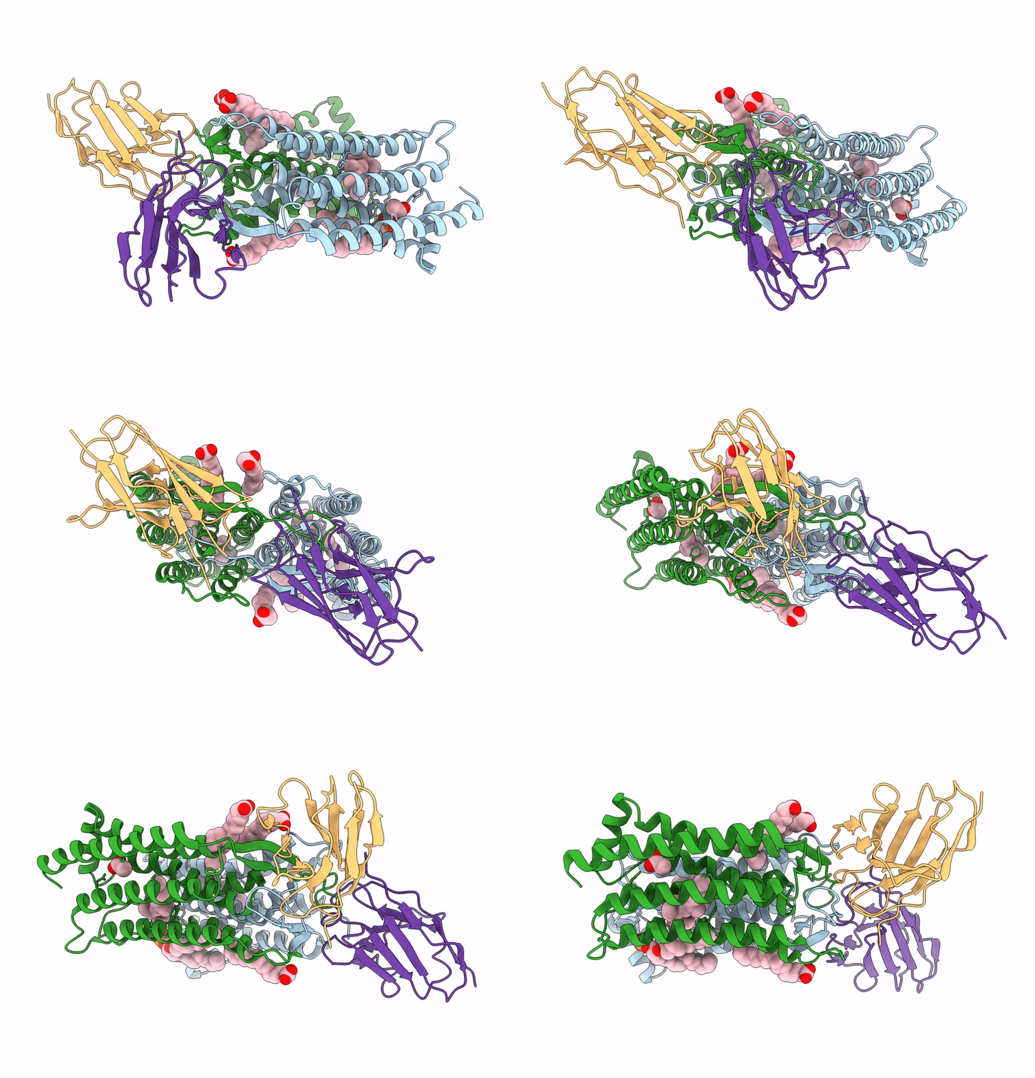
9LJJ image
Entry Detail
PDB ID:
9LJJ
EMDB ID:
Title:
cryo-EM structure of a nanobody bound heliorhodopsin
Biological Source:
Host Organism:
PDB Version:
Deposition Date:
2025-01-15
Release Date:
2025-02-12
Method Details:
Experimental Method:
Resolution:
3.23 Å
Aggregation State:
PARTICLE
Reconstruction Method:
SINGLE PARTICLE
Macromolecular Entities
Polymer Type:polypeptide(L)
Description:Heliorhodopsin
Chain IDs:A, B
Chain Length:256
Number of Molecules:2
Biological Source:Escherichia coli
Polymer Type:polypeptide(L)
Description:nanobody B4
Chain IDs:C (auth: M), D (auth: N)
Chain Length:122
Number of Molecules:2
Biological Source:Vicugna pacos
Primary Citation
Cryo-EM structure of a nanobody-bound heliorhodopsin.
Biochem.Biophys.Res.Commun. 750 151398 151398 (2025)
PMID: 39889627 DOI: 10.1016/j.bbrc.2025.151398

Abstact

Heliorhodopsins (HeRs) represent a distinct class of microbial rhodopsins (MRs) with an inverted membrane topology compared to other MRs. Previous structural studies have shown that HeRs lack a proton acceptor residue, and protons are never released from the protein. In this study, we present the cryo-electron microscopy (cryo-EM) structure of HeR bound to a nanobody. The structure reveals an acetate-like molecule in the Schiff base cavity (SBC) on the intracellular side of HeR under neutral condition. Structural comparisons and analyses suggest that the acetate molecule may function as a proton acceptor for the protonated retinal Schiff base (RSB) and act as a mediator for the intramolecular signaling transduction in HeR during light stimulation. These structural insights shed new light on the mechanism and function of HeR.

Legend

Protein

Chemical

Disease

Primary Citation of related structures