9LJ8 image
Entry Detail
PDB ID:
9LJ8
EMDB ID:
Keywords:
Title:
Tail structure of bacteriophage Mu in contracted state
Biological Source:
Source Organism:
PDB Version:
Deposition Date:
2025-01-14
Release Date:
2025-02-19
Method Details:
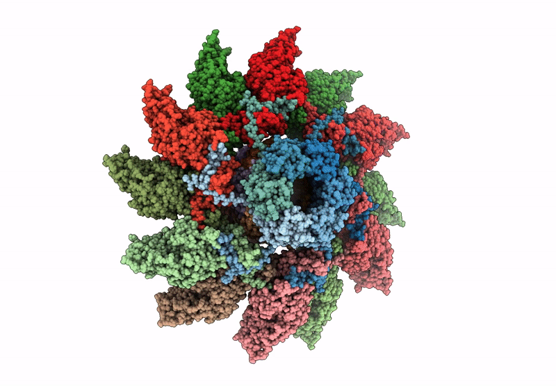
Experimental Method:
Resolution:
3.80 Å
Aggregation State:
PARTICLE
Reconstruction Method:
SINGLE PARTICLE
Macromolecular Entities
Polymer Type:polypeptide(L)
Description:Probable tail terminator protein
Chain IDs:A, F (auth: B), G (auth: C), H (auth: D), I (auth: E), J (auth: F)
Chain Length:118
Number of Molecules:6
Biological Source:Escherichia phage Mu
Polymer Type:polypeptide(L)
Description:Tail sheath protein
Chain IDs:D (auth: M), E (auth: G), U (auth: H), V (auth: I), W (auth: J), X (auth: K), Y (auth: L), Z (auth: N), AA (auth: O), BA (auth: P), CA (auth: Q), DA (auth: R)
Chain Length:495
Number of Molecules:12
Biological Source:Escherichia phage Mu
Polymer Type:polypeptide(L)
Description:Tail tube protein
Chain IDs:B (auth: a), C (auth: S), K (auth: T), L (auth: U), M (auth: V), N (auth: W), O (auth: X), P (auth: Y), Q (auth: Z), R (auth: b), S (auth: c), T (auth: d)
Chain Length:118
Number of Molecules:12
Biological Source:Escherichia phage Mu
Ligand Molecules
Primary Citation
In situ structures of the contractile nanomachine myophage Mu in both its extended and contracted states.
J.Virol. 99 e0205624 e0205624 (2025)
PMID: 39992138 DOI: 10.1128/jvi.02056-24

Abstact

UNLABELLED Myophage Mu is a representative of contractile nanomachines with a simple tail baseplate. It has the capacity to infect a range of intestinal bacteria and has extensive applications in genetic engineering research. Nevertheless, a comprehensive understanding of the entire structure and contractile mechanisms of Mu remains elusive. Using cryo-electron microscopy (cryo-EM), we resolved the asymmetric structures of Mu in both its extended and contracted states, the latter of which lacked the tail baseplate, at near-atomic resolutions. We built the atomic models for the extended Mu, encompassing the head, the connector complex, the tail, and the simple baseplate. It is noteworthy that we identified the position and structure of the tail tube initiator protein gp43 (referred to as the DNA circularization protein). The protein gp43 plays a crucial role not only in the baseplate assembly and DNA circularization but also in stabilizing the wedge-hub connection and mediating tail contraction. Except for the baseplate structure, the structural comparison of Mu in its extended and contracted states revealed that only the tail sheath protein gp39 and the C-terminus of the tail terminator protein gp37 undergo notable conformational changes to accommodate the tail contraction, whereas the remaining protein components remained unchanged. Our structures exhibited conserved properties among the majority of myophages, thereby providing valuable insights into the contraction mechanisms across myophages and contractile injection systems (CISs). IMPORTANCE Despite extensive study, the asymmetric structures of phage Mu, a highly effective transposable myophage, remain unknown. In this study, we present the high-resolution structures of Mu in both its extended and contracted states. The comparison of the two structures allows for the illustration of detailed conformational changes of the head-to-tail complex during the process of tail contraction. The contraction mechanism of Mu is highly conserved and widely adapted to all contractile nanomachines that share common structural features with Mu.

Legend

Protein

Chemical

Disease

Primary Citation of related structures