9LIP image
Deposition Date 2025-01-14
Release Date 2025-12-24
Last Version Date 2025-12-24
Entry Detail
PDB ID:
9LIP
Keywords:
Title:
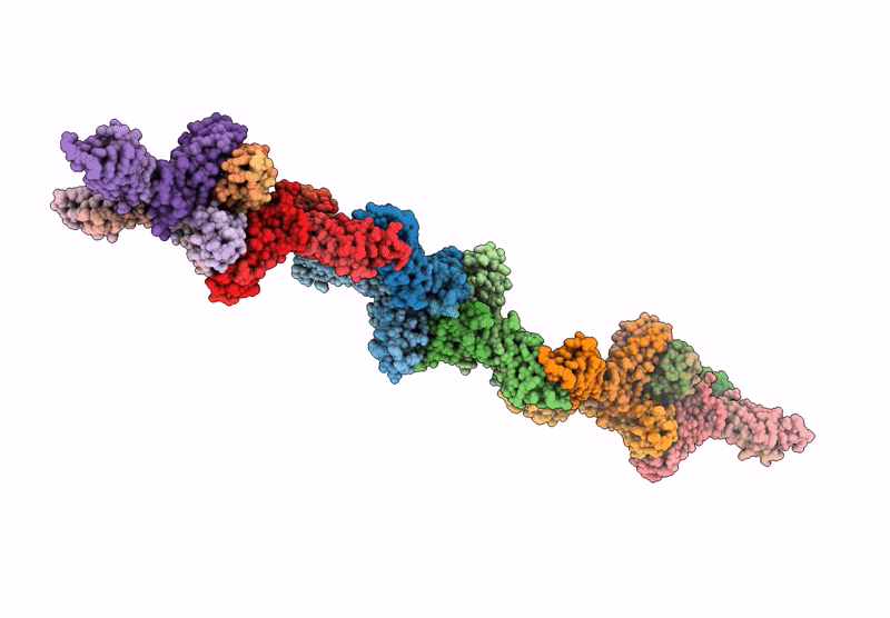
The cryo-EM structure of the native PMEL fibril lamella
Biological Source:
Source Organism:
Homo sapiens (Taxon ID: 9606)
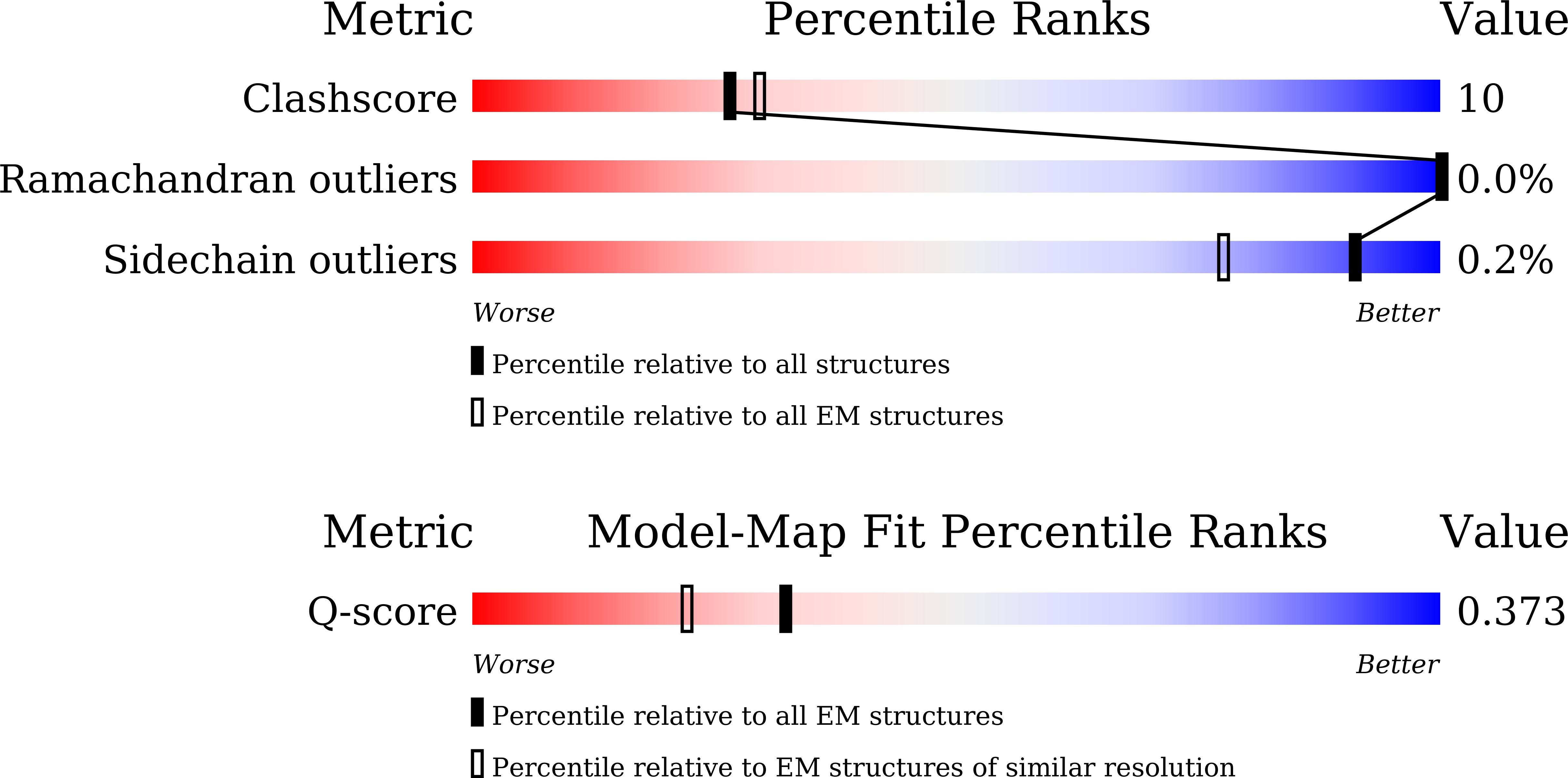
Method Details:
Experimental Method:
Resolution:
3.48 Å
Aggregation State:
FILAMENT
Reconstruction Method:
SINGLE PARTICLE
Macromolecular Entities
Structures with similar UniProt ID
Protein Blast
Polymer Type:polypeptide(L)
Molecule:M-alpha
Gene (Uniprot):PMEL
Chain IDs:A (auth: Q), E (auth: U), I (auth: A), M (auth: E), Q (auth: I), U (auth: M), Y (auth: g), CA (auth: k), GA (auth: Y), KA (auth: c), OA (auth: o), SA (auth: s)
Chain Length:24
Number of Molecules:12
Biological Source:Homo sapiens
Structures with similar UniProt ID
Protein Blast
Polymer Type:polypeptide(L)
Molecule:M-alpha
Gene (Uniprot):PMEL
Chain IDs:B (auth: R), F (auth: V), J (auth: B), N (auth: F), R (auth: J), V (auth: N), Z (auth: h), DA (auth: l), HA (auth: Z), LA (auth: d), PA (auth: p), TA (auth: t)
Chain Length:17
Number of Molecules:12
Biological Source:Homo sapiens
Structures with similar UniProt ID
Protein Blast
Polymer Type:polypeptide(L)
Molecule:M-alpha
Gene (Uniprot):PMEL
Chain IDs:C (auth: S), G (auth: W), K (auth: C), O (auth: G), S (auth: K), W (auth: O), AA (auth: i), EA (auth: m), IA (auth: a), MA (auth: e), QA (auth: q), UA (auth: u)
Chain Length:24
Number of Molecules:12
Biological Source:Homo sapiens
Structures with similar UniProt ID
Protein Blast
Polymer Type:polypeptide(L)
Molecule:M-beta
Gene (Uniprot):PMEL
Chain IDs:D (auth: T), H (auth: X), L (auth: D), P (auth: H), T (auth: L), X (auth: P), BA (auth: j), FA (auth: n), JA (auth: b), NA (auth: f), RA (auth: r), VA (auth: v)
Chain Length:17
Number of Molecules:12
Biological Source:Homo sapiens
Ligand Molecules
Primary Citation
Atomic structure and in situ visualization of native PMEL lamellae in melanosomes.
Nat Commun 16 10300 10300 (2025)
PMID: 41271718 DOI: 10.1038/s41467-025-65221-0

Abstact

Melanin synthesis within melanosomes critically depends on the fibrillar architecture formed by the pigment cell-specific protein PMEL. Although PMEL fibrils have historically been classified as functional amyloids, their native supramolecular organization in situ and detailed molecular architecture have remained unresolved. In this study, we combine in situ cryo-electron tomography (cryo-ET) and cryo-electron microscopy (cryo-EM) to elucidate the native structural organization of PMEL fibrils within human melanosomes from both patient melanoma tissues and melanocyte cell lines. We demonstrate that PMEL does not form conventional isolated amyloid fibrils, but rather assembles into highly organized lamellar sheets consisting of laterally aligned fibrils interconnected by flexible linker regions. Cryo-EM structures reveal a distinctive butterfly-shaped fibril unit composed of multiple structured domains, including both the proteolytically cleaved Mα and Mβ fragments of PMEL, which assemble into a amyloid-like β-sheet arrangement. Notably, we identify intrinsically disordered regions critical for lamellar assembly and curvature and verify key glycosylation modifications in the structure. This architecture distinguishes PMEL fibrils from canonical amyloids and elucidates the molecular basis underlying melanosome integrity and pigmentation. Moreover, our work provides molecular insights relevant for pigmentation disorders and PMEL-associated diseases, including melanoma.

Legend

Protein

Chemical

Disease

Primary Citation of related structures
Feedback Form
Name
Email
Institute
Feedback