9LFL image
Deposition Date 2025-01-08
Release Date 2025-08-13
Last Version Date 2025-09-17
Entry Detail
PDB ID:
9LFL
Keywords:
Title:
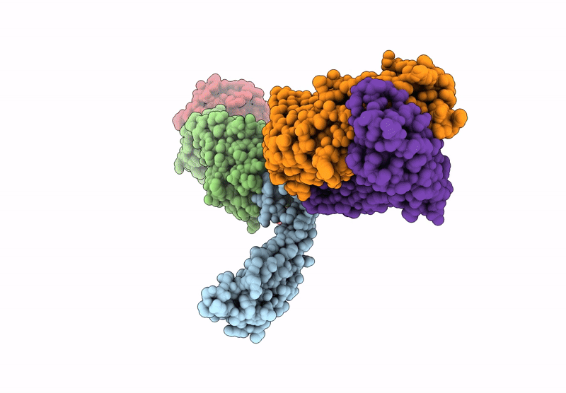
Cryo-EM structure of linker-extended biparatopic antibody BA1-GP4 in complex with TNFR2
Biological Source:
Source Organism:
Homo sapiens (Taxon ID: 9606)
Mus musculus (Taxon ID: 10090)
Host Organism:
Method Details:
Experimental Method:
Resolution:
3.73 Å
Aggregation State:
PARTICLE
Reconstruction Method:
SINGLE PARTICLE
Macromolecular Entities
Structures with similar UniProt ID
Protein Blast
Polymer Type:polypeptide(L)
Molecule:Tumor necrosis factor receptor superfamily member 1B
Gene (Uniprot):TNFRSF1B
Mutagens:A490V
Chain IDs:A
Chain Length:152
Number of Molecules:1
Biological Source:Homo sapiens
Polymer Type:polypeptide(L)
Molecule:TR92 heavy chain
Chain IDs:B
Chain Length:221
Number of Molecules:1
Biological Source:Mus musculus
Polymer Type:polypeptide(L)
Molecule:TR92 light chain
Chain IDs:C
Chain Length:211
Number of Molecules:1
Biological Source:Mus musculus
Polymer Type:polypeptide(L)
Molecule:TR96 heavy chain
Chain IDs:D
Chain Length:219
Number of Molecules:1
Biological Source:Mus musculus
Polymer Type:polypeptide(L)
Molecule:TR96 light chain
Chain IDs:E
Chain Length:210
Number of Molecules:1
Biological Source:Mus musculus
Ligand Molecules
Primary Citation
Conversion of an agonistic anti-TNFR2 biparatopic antibody into an antagonist by insertion of peptide linkers into the hinge region.
J.Biol.Chem. 301 110548 110548 (2025)
PMID: 40752574 DOI: 10.1016/j.jbc.2025.110548

Abstact

Biparatopic antibodies (BpAbs) bind two different antigen epitopes to form characteristic immunocomplexes. Many BpAbs have been developed for enhanced cross-linking to induce signal transduction or cell internalization, whereas few were reported with smaller immunocomplexes to suppress unwanted signaling. Here, we developed a strategy to induce 1:1 immunocomplex formation to maximize antagonistic function. Various peptide linkers were introduced into the hinge regions of IgG-like agonist BpAbs against tumor necrosis factor receptor 2. Loss of crosslinking activity was observed for one BpAb, allowing the conversion of its function from an agonist to an antagonist. However, cross-linking activity was retained for another agonist BpAb, which binds to a different epitope pair. In a combined analysis of cryo-electron microscopy and coarse-grained molecular dynamics simulations, effect of epitope combination on the stability of 1:1 complexes was observed. These results lead to an understanding of the mechanism and design of BpAbs to adopt a 1:1-binding mode.

Legend

Protein

Chemical

Disease

Primary Citation of related structures
Feedback Form
Name
Email
Institute
Feedback