9L9T image
Deposition Date 2024-12-31
Release Date 2025-11-05
Last Version Date 2025-11-05
Entry Detail
PDB ID:
9L9T
Title:
Crystal structure of a coronaviral M protein in complex with a C-terminal peptide of the N protein
Biological Source:
Source Organism:
Host Organism:
Method Details:
Experimental Method:
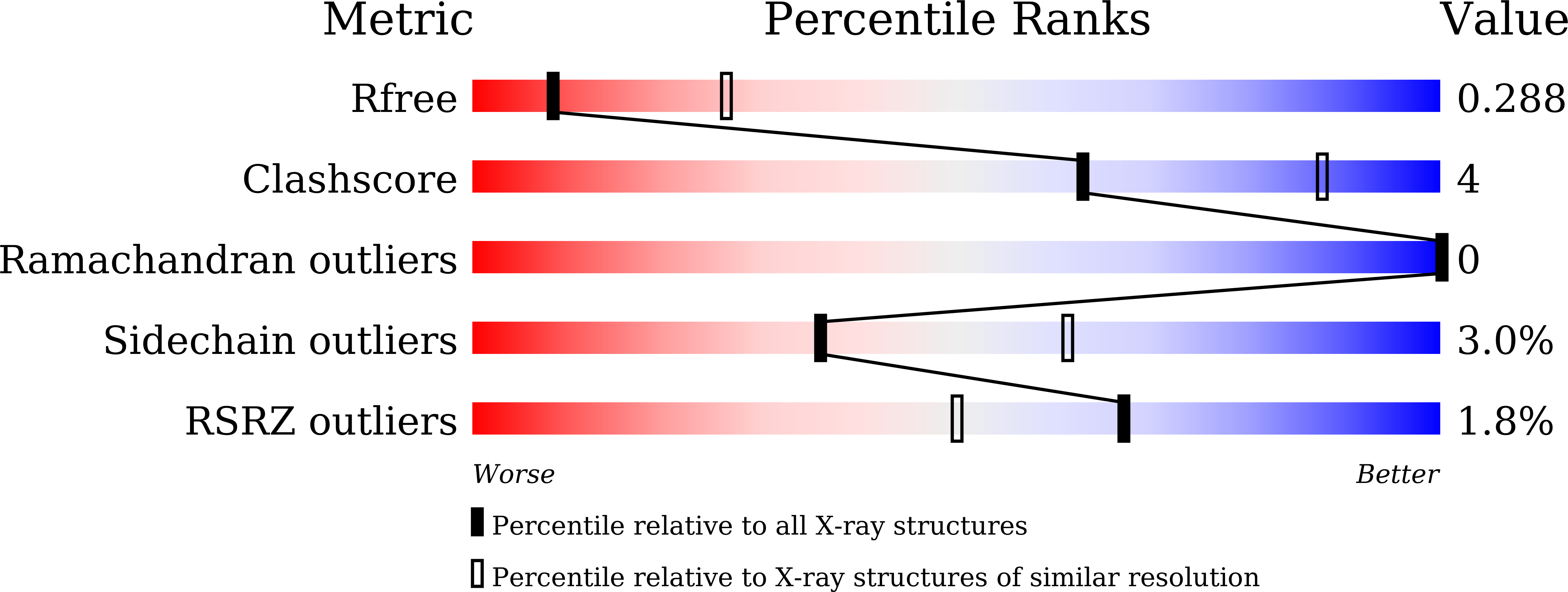
Resolution:
3.14 Å
R-Value Free:
0.29
R-Value Work:
0.25
R-Value Observed:
0.25
Space Group:
C 2 2 21
Macromolecular Entities
Structures with similar UniProt ID
Protein Blast
Polymer Type:polypeptide(L)
Molecule:Membrane protein
Gene (Uniprot):M
Chain IDs:A, B
Chain Length:220
Number of Molecules:2
Biological Source:Pipistrellus bat coronavirus HKU5
Polymer Type:polypeptide(L)
Molecule:N protein peptide poly-Ala model
Chain IDs:C, D
Chain Length:5
Number of Molecules:2
Biological Source:Pipistrellus bat coronavirus HKU5
Ligand Molecules
Primary Citation
Binding of an N protein peptide to M protein of a bat coronavirus.
J.Struct.Biol. 217 108234 108234 (2025)
PMID: 40664277 DOI: 10.1016/j.jsb.2025.108234

Abstact

The interaction between the membrane (M) protein and the nucleocapsid (N) protein of coronaviruses plays a crucial role in virus assembly and morphogenesis. Previous studies indicate that one M-N interaction occurs between M protein and the carboxy-terminus of N protein. However, the mechanistic details of M-N interactions remain unclear. Here, we present a complex structure of an N protein carboxy-terminal peptide bound to M protein from Pipistrellus bat coronavirus HKU5. The structure shows that the M-N peptide binding site includes a "horizontal" groove located between the carboxy-terminal domain and the transmembrane domain of M protein. Combined with molecular docking and binding analysis, our results provide structural insight into the binding mechanism between M and N proteins of a coronavirus.

Legend

Protein

Chemical

Disease

Primary Citation of related structures