9L2N image
Deposition Date 2024-12-17
Release Date 2025-07-02
Last Version Date 2025-07-30
Entry Detail
PDB ID:
9L2N
Title:
Crystal structure of Cytochalasin D bound to a filamentous conformation actin
Biological Source:
Source Organism:
Host Organism:
Method Details:
Experimental Method:
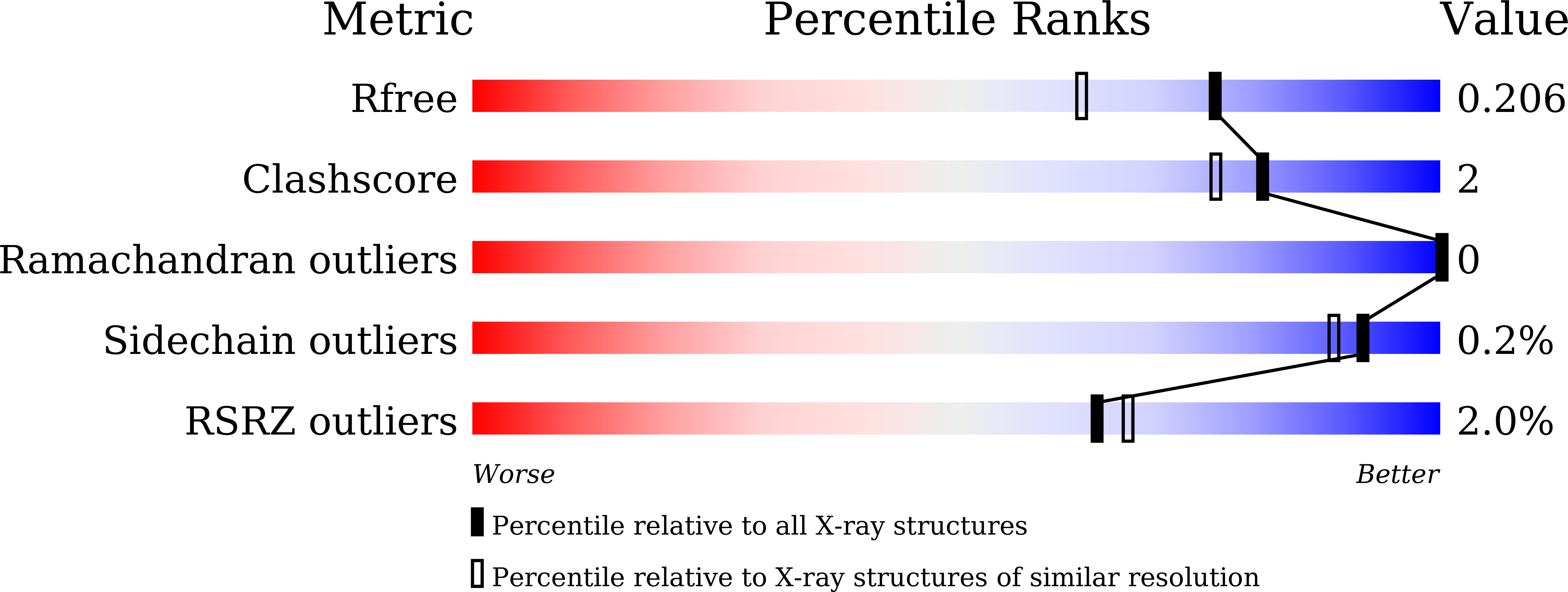
Resolution:
1.70 Å
R-Value Free:
0.20
R-Value Work:
0.17
R-Value Observed:
0.17
Space Group:
P 21 21 21
Macromolecular Entities
Structures with similar UniProt ID
Protein Blast
Polymer Type:polypeptide(L)
Molecule:Actin, alpha skeletal muscle
Gene (Uniprot):ACTA1
Chain IDs:A
Chain Length:375
Number of Molecules:1
Biological Source:Gallus gallus
Structures with similar UniProt ID
Protein Blast
Polymer Type:polypeptide(L)
Molecule:Actin-binding protein fragmin P
Mutagens:N13A
Chain IDs:B
Chain Length:162
Number of Molecules:1
Biological Source:Physarum polycephalum
Modified Residue
Compound ID Chain ID Parent Comp ID Details 2D Image
HIC A HIS modified residue
Primary Citation
Microscopic and structural observations of actin filament capping and severing by cytochalasin D.
Proc.Natl.Acad.Sci.USA 122 e2502164122 e2502164122 (2025)
PMID: 40658853 DOI: 10.1073/pnas.2502164122

Abstact

Cytochalasin D (CytoD), a widely used actin inhibitor, is typically employed in cell studies as a simple barbed end capper. However, accumulating evidence suggests broader effects on actin dynamics. We addressed this by observing single actin filaments using total internal reflection fluorescence microscopy. Our depolymerization assay confirmed that, at nanomolar concentrations, CytoD tightly caps barbed ends. The K1/2 for inhibition was 4.1 nM, consistent with previous bulk measurements, and our approach revealed a capping duration of ~2 min. In polymerization assays, nanomolar CytoD completely suppressed barbed end elongation. Interestingly, at subnanomolar concentrations, CytoD caps barbed ends only transiently, rapidly associating and dissociating under both polymerizing and depolymerizing conditions. We interpreted this contradictory behavior as arising from differences in binding modes: capping one strand (fast dissociation) or both strands (slow dissociation). CytoD severs actin filaments at micromolar levels, a concentration range commonly used in cell biological studies. Although the severing rate is slower than cofilin, its higher frequency leads to filament fragmentation. Severing activity was suppressed by inorganic phosphate or cofilin. Our crystal structure of CytoD bound to filamentous conformation (F-form) actin showed that CytoD fits better in the hydrophobic cleft of F-form actin than monomeric conformation actin, explaining its preference for barbed end subunits. CytoD prevents barbed end depolymerization by stabilizing the terminal subunits in the F-form, which is supported by our MD simulations. These findings reveal the molecular mechanisms by which CytoD modulates actin dynamics and highlight the need for careful dosage control when treating cells with CytoD.

Legend

Protein

Chemical

Disease

Primary Citation of related structures