9KUE image
Deposition Date 2024-12-03
Release Date 2025-04-23
Last Version Date 2025-09-10
Entry Detail
PDB ID:
9KUE
Title:
Crystal structure of the soluble green pigment protein from Tettigonia cantans
Biological Source:
Source Organism:
Method Details:
Experimental Method:
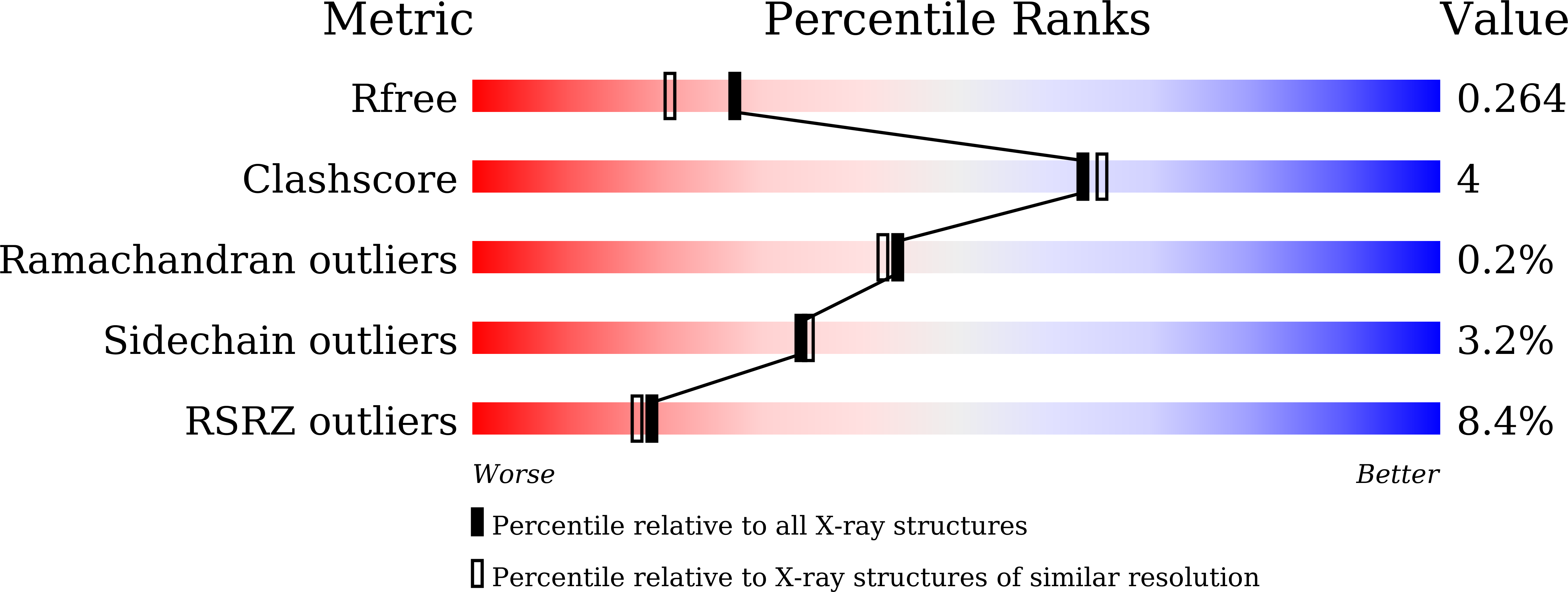
Resolution:
1.99 Å
R-Value Free:
0.26
R-Value Work:
0.22
R-Value Observed:
0.22
Space Group:
P 21 21 21
Macromolecular Entities
Polymer Type:polypeptide(L)
Molecule:Dibilinoxanthinin (DBXN)
Chain IDs:A, D
Chain Length:114
Number of Molecules:2
Biological Source:Tettigonia cantans
Polymer Type:polypeptide(L)
Molecule:Dibilinoxanthinin (DBXN)
Chain IDs:B, E
Chain Length:67
Number of Molecules:2
Biological Source:Tettigonia cantans
Polymer Type:polypeptide(L)
Molecule:Dibilinoxanthinin (DBXN)
Chain IDs:C, F
Chain Length:172
Number of Molecules:2
Biological Source:Tettigonia cantans
Primary Citation
A green dichromophoric protein enabling foliage mimicry in arthropods.
Proc.Natl.Acad.Sci.USA 122 e2502567122 e2502567122 (2025)
PMID: 40424164 DOI: 10.1073/pnas.2502567122

Abstact

Molecular mechanisms underlying the green insect camouflage have puzzled researchers for over a century. Here, we isolated and identified a green water-soluble protein from the integument of bush-cricket Tettigonia cantans. De novo sequencing and cloning revealed a severely fragmented form of vitellogenins, ubiquitous and multifunctional, but still largely enigmatic glycolipoproteins essential for embryonic development and lacking structural characterization. The distinctive color of the identified chromoprotein results from binding of a remarkable combination of farnesylated bilins (recently identified, tentative heme A catabolites) and xanthophylls, which commensurably absorb light in the 600 to 700 nm and 400 to 550 nm spectral regions and thereby produce a hue that perfectly mimics foliage. The high-resolution crystal structure of this unique ~80 kDa dichromophoric protein, which we named "dibilinoxanthinin" (DBXN), revealed two DBXN protomers, each consisting of three polypeptides, with a novel fold enclosing a large hydrophobic cavity that accommodates two bilins, two luteins, and four phosphatidylcholines, all anchored by hydrogen bonds and giving DBXN unique biochemical and optical properties. Among the green insects tested, some contained yellow and blue chromophores in separate fractions, while others had green proteins similar to DBXN, although not necessarily of the same size. Surprisingly, we isolated and identified a larger vitellogenin proteoform with DBXN-like absorption, from the green huntsman spider Micrommata virescens. These data illustrate striking variations in the DBXN-related pigmentation mechanism among different green arthropods and suggest that vitellogenins may have undergone neofunctionalization, reflecting their potential for functional diversification.

Legend

Protein

Chemical

Disease

Primary Citation of related structures