9KME image
Deposition Date 2024-11-15
Release Date 2025-06-04
Last Version Date 2025-07-02
Entry Detail
PDB ID:
9KME
Keywords:
Title:
Crystal structure of soluble bacteriorhodopsin NeuroBR_A
Biological Source:
Source Organism:
Host Organism:
Method Details:
Experimental Method:
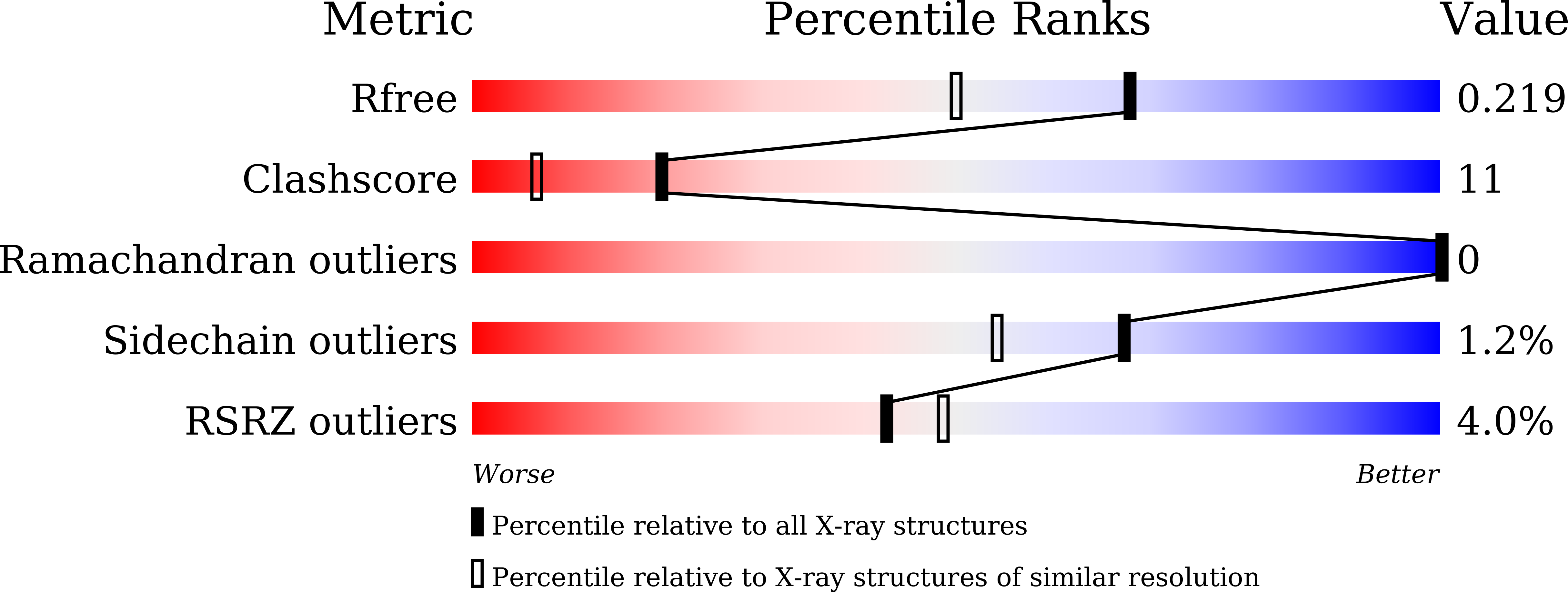
Resolution:
1.76 Å
R-Value Free:
0.21
R-Value Work:
0.17
R-Value Observed:
0.17
Space Group:
P 21 21 21
Macromolecular Entities
Polymer Type:polypeptide(L)
Molecule:soluble bacteriorhodopsin
Chain IDs:A, B, C, D
Chain Length:237
Number of Molecules:4
Biological Source:synthetic construct
Primary Citation

Abstact

Studies and applications of membrane proteins remain challenging due to the requirement of maintaining them in a lipid membrane or a membrane mimic. Modern machine learning-based protein engineering methods offer a possibility of generating soluble analogs of membrane proteins that retain the active site structure and ligand-binding properties; however, clear examples are currently missing. Here, we report successful engineering of proteins dubbed NeuroBRs that mimic the active site (retinal-binding pocket) of bacteriorhodopsin, a light-driven proton pump and well-studied model membrane protein. NeuroBRs are soluble and stable, bind retinal and exhibit photocycles under illumination. The crystallographic structure of NeuroBR_A, determined at anisotropic resolution reaching 1.76 Å, reveals an excellently conserved chromophore binding pocket and tertiary structure. Thus, NeuroBRs are promising microbial rhodopsin mimics for studying retinal photochemistry and potential soluble effector modules for optogenetic tools. Overall, our results highlight the power of modern protein engineering approaches and pave the way towards wider development of molecular tools derived from membrane proteins.

Legend

Protein

Chemical

Disease

Primary Citation of related structures