9KLT image
Deposition Date 2024-11-15
Release Date 2026-01-28
Last Version Date 2026-01-28
Entry Detail
PDB ID:
9KLT
Keywords:
Title:
Crystal structure of a Streptococcal protein G B1 mutant
Biological Source:
Source Organism(s):
Expression System(s):
Method Details:
Experimental Method:
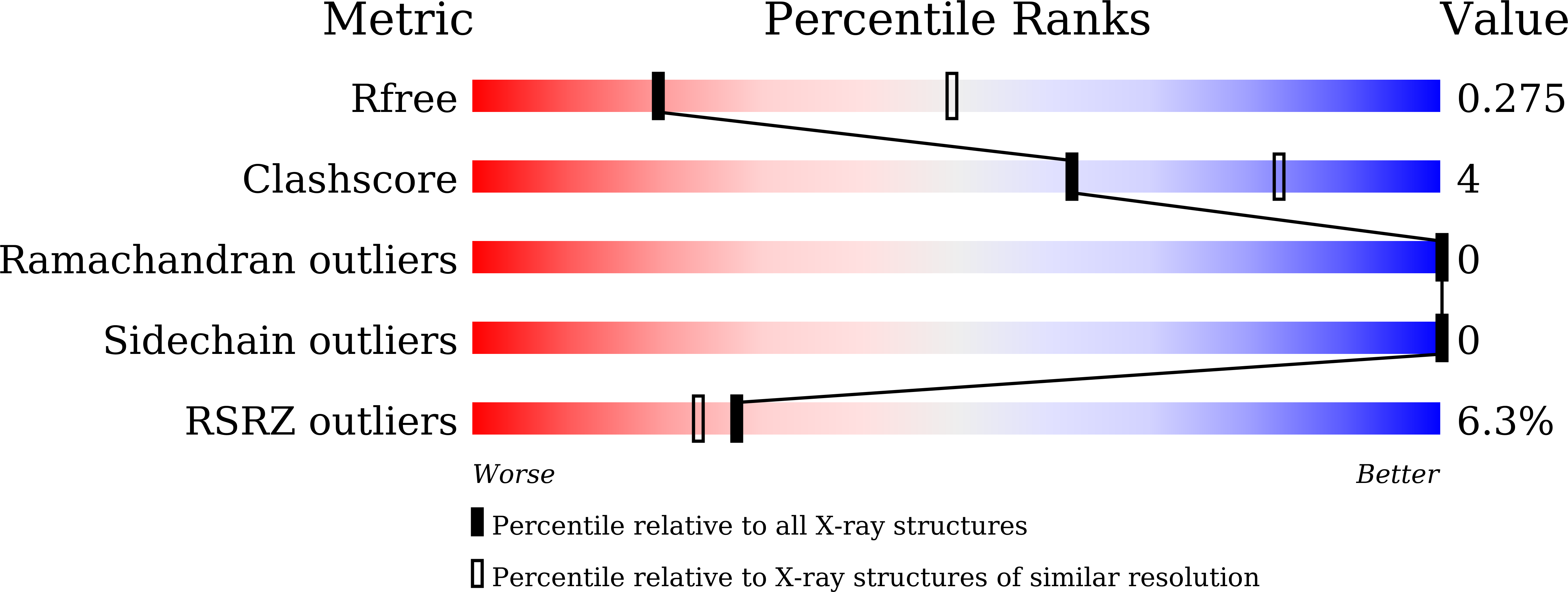
Resolution:
2.88 Å
R-Value Free:
0.27
R-Value Work:
0.22
R-Value Observed:
0.23
Space Group:
I 21 21 21
Macromolecular Entities
Structures with similar UniProt ID
Protein Blast
Polymer Type:polypeptide(L)
Molecule:Protein LG
Mutagens:D365E/N365H/D375P/A376E
Chain IDs:A
Chain Length:66
Number of Molecules:1
Biological Source:Finegoldia magna
Ligand Molecules
Primary Citation
Regulating the orientation of homobivalent small binders through 3D domain-swapping design.
J.Biol.Chem. 302 111053 111053 (2025)
PMID: 41391770 DOI: 10.1016/j.jbc.2025.111053

Abstact

Homobivalent small binders represent bioactive dimeric proteins constructed by linking nanobodies or other small binders. As binding domain orientation in these dimeric binders affects binding affinity, expanding the orientation regulation techniques is crucial for optimizing their performance. Here, we report a 3D domain-swapping design as a genetic engineering approach to regulate the orientation of homobivalent small binders. We aimed to regulate B1 domain orientation of protein G as a model binder. By inserting single cysteine-containing polyproline sequences into loops located at the C- and N-terminal sides of the tertiary structure, we successfully designed 3D domain-swapped dimers with tail-to-tail and head-to-head orientations. Notably, the head-to-head-oriented dimer demonstrated potential as a functional homobivalent small binder. Our results highlight the applicability of the 3D domain-swapping design for regulating the orientation of homobivalent small binders and establish a foundation for developing 3D domain-swapped dimers.

Legend

Protein

Chemical

Disease

Primary Citation of related structures
Feedback Form
Name
Email
Institute
Feedback