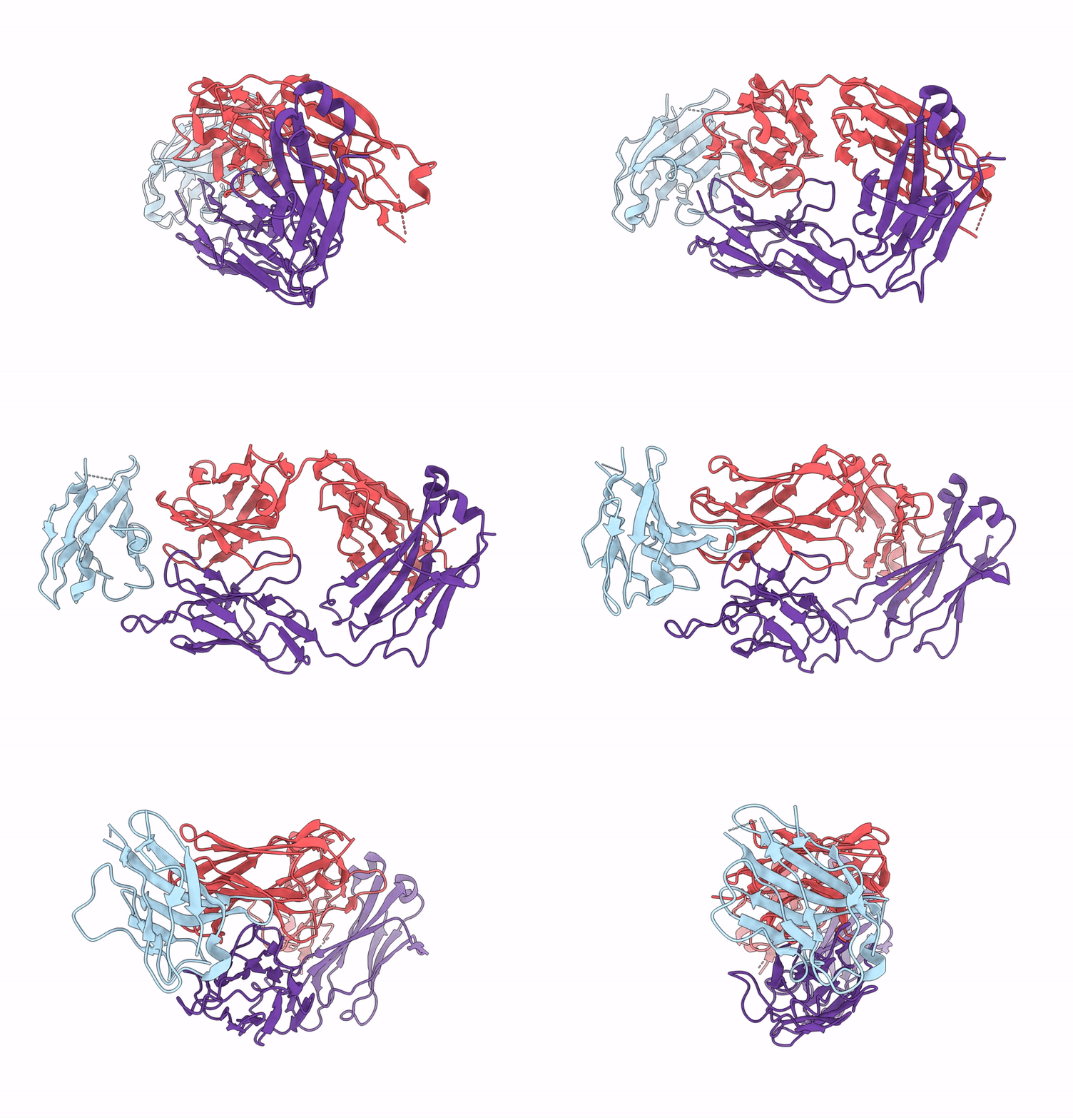
9KKJ image
Deposition Date 2024-11-13
Release Date 2025-11-19
Last Version Date 2025-11-19
Entry Detail
PDB ID:
9KKJ
Keywords:
Title:
Structure of Nectin-4 D1 domain in complex with the Fab fragment of 9MW2821 mAb
Biological Source:
Source Organism:
Mus musculus (Taxon ID: 10090)
Homo sapiens (Taxon ID: 9606)
Host Organism:
Method Details:
Experimental Method:
Resolution:
3.26 Å
Aggregation State:
PARTICLE
Reconstruction Method:
SINGLE PARTICLE
Macromolecular Entities
Structures with similar UniProt ID
Protein Blast
Polymer Type:polypeptide(L)
Molecule:Nectin-4
Gene (Uniprot):NECTIN4
Chain IDs:C (auth: A)
Chain Length:114
Number of Molecules:1
Biological Source:Homo sapiens
Polymer Type:polypeptide(L)
Molecule:9MW2821 mAb Fab Heavy Chain
Chain IDs:B (auth: H)
Chain Length:220
Number of Molecules:1
Biological Source:Mus musculus
Polymer Type:polypeptide(L)
Molecule:9MW2821 mAb Fab Light chain
Chain IDs:A (auth: L)
Chain Length:218
Number of Molecules:1
Biological Source:Mus musculus
Ligand Molecules
Primary Citation
Structural basis of nectin-4 recognition by the antibody-drug conjugate 9MW2821.
J.Biol.Chem. ? 110816 110816 (2025)
PMID: 41101504 DOI: 10.1016/j.jbc.2025.110816

Abstact

Nectin-4, a membrane protein highly expressed in multiple solid tumors, has become an attractive target for antibody-drug conjugate development. We designed and developed 9MW2821, an anti-nectin-4 antibody-drug conjugate with an enzymatically cleavable valine-citrulline linker and monomethyl auristatin E as the payload. Although 9MW2821 has shown good efficacy and safety in multiple solid tumors in clinical trials, the interaction between 9MW2821 and nectin-4 at the molecular level remains unclear. In this study, we solved the structure of the antigen-binding fragment of 9MW2821 in complex with nectin-4 at a resolution of 3.26 Å using single-particle cryo-EM. The structure shows that 9MW2821 binds the front β-sheet of nectin-4 D1 through three complementarity-determining region loops from the heavy chain and two complementarity-determining region loops from the light chain. The binding involves extensive hydrogen bonds and hydrophobic interactions. The buried surface area is more than 1600 Å2. Mutagenesis studies revealed that four residues (Q77, E78, H83, and E95) of nectin-4 contributed significantly to the binding of 9MW2821 monoclonal antibody (mAb). The structure also shows that 9MW2821 mAb blocks nectin-4 homodimer formation by competing with the nectin-4 partner D1 domain for part of the nectin-4 surface area. Furthermore, 9MW2821 mAb prevents nectin-4 from interacting with nectin-1 and T-cell immunoglobulin and immunoreceptor tyrosine-based inhibitory motif domain, enhancing the activation of NK cells. Based on the structure of the nectin-4 D1-9MW2821 antigen-binding fragment complex resolved in this study, we have elucidated the molecular mechanism of 9MW2821 in cancer therapy. The findings provide a basis for optimizing future mAbs targeting nectin-4.

Legend

Protein

Chemical

Disease

Primary Citation of related structures
Feedback Form
Name
Email
Institute
Feedback