9KHS image
Deposition Date 2024-11-11
Release Date 2025-07-30
Last Version Date 2025-08-27
Entry Detail
PDB ID:
9KHS
Keywords:
Title:
Cryo-EM structure of Ufd2/Ubc4-Ub in complex with K29-linked diUb (monomeric conformation)
Biological Source:
Host Organism:
Method Details:
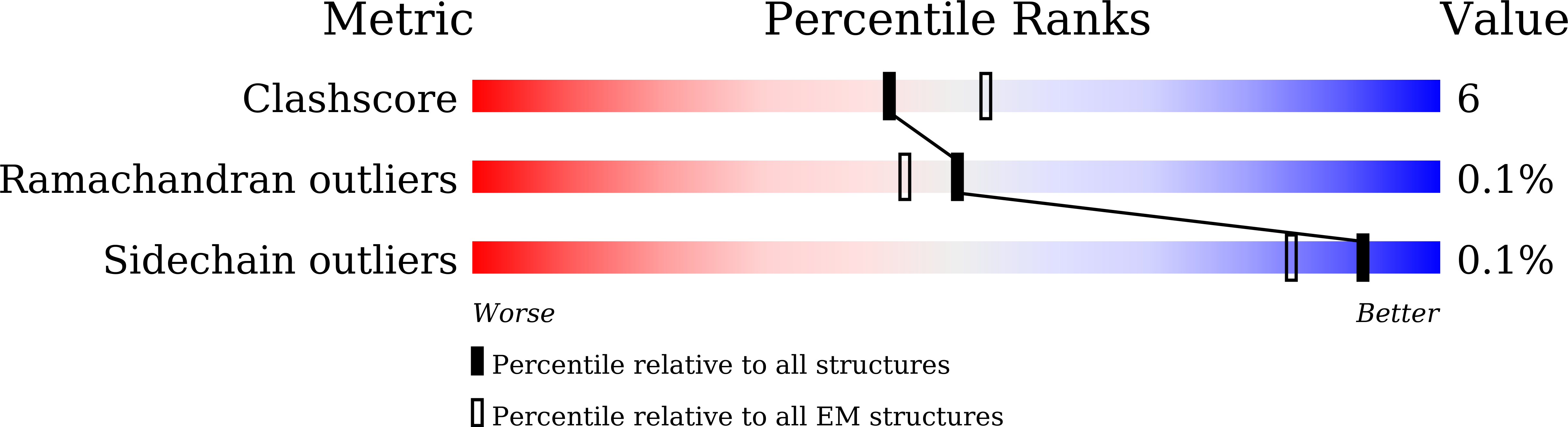
Experimental Method:
Resolution:
4.31 Å
Aggregation State:
PARTICLE
Reconstruction Method:
SINGLE PARTICLE
Macromolecular Entities
Structures with similar UniProt ID
Protein Blast
Polymer Type:polypeptide(L)
Molecule:E4 ubiquitin-protein ligase UFD2
Gene (Uniprot):UFD2
Chain IDs:E (auth: A)
Chain Length:961
Number of Molecules:1
Biological Source:Saccharomyces cerevisiae (strain ATCC 204508 / S288c)
Structures with similar UniProt ID
Protein Blast
Polymer Type:polypeptide(L)
Molecule:Ubiquitin-conjugating enzyme E2 4
Gene (Uniprot):UBC4
Mutagens:C22S/C102S
Chain IDs:A (auth: B)
Chain Length:148
Number of Molecules:1
Biological Source:Saccharomyces cerevisiae (strain ATCC 204508 / S288c)
Structures with similar UniProt ID
Protein Blast
Polymer Type:polypeptide(L)
Molecule:Ubiquitin
Gene (Uniprot):RPS27A
Chain IDs:D (auth: C)
Chain Length:76
Number of Molecules:1
Biological Source:Homo sapiens
Structures with similar UniProt ID
Protein Blast
Polymer Type:polypeptide(L)
Molecule:Polyubiquitin-C
Gene (Uniprot):UBC
Mutagens:K48C
Chain IDs:B (auth: D)
Chain Length:77
Number of Molecules:1
Biological Source:Homo sapiens
Structures with similar UniProt ID
Protein Blast
Polymer Type:polypeptide(L)
Molecule:Polyubiquitin-C
Gene (Uniprot):UBC
Mutagens:K29R
Chain IDs:C (auth: E)
Chain Length:76
Number of Molecules:1
Biological Source:Homo sapiens
Ligand Molecules
Primary Citation
Structural basis for E4 enzyme Ufd2-catalyzed K48/K29 branched ubiquitin chains.
Nat.Chem.Biol. ? ? ? (2025)
PMID: 40817136 DOI: 10.1038/s41589-025-01985-2

Abstact

E4 enzymes amplify and remodel ubiquitin chain signals beyond the conventional E1-E2-E3 cascade. The first identified E4 enzyme Ufd2 preferentially catalyzes K48/K29 branched ubiquitin chains, yet the structural mechanism remains unknown. Here, we combined chemical biology and cryo-electron microscopy to visualize stable intermediates in Ufd2 loading ubiquitin at K48 of proximal ubiquitin on K29-linked di- and triubiquitin. Our data reveal that the core region of Ufd2 functions as an unprecedented K29 diubiquitin binding domain, interacting extensively with proximal and distal ubiquitin, which orients the K48 site of proximal ubiquitin toward the active site of Ubc4, facilitating K48/K29 branched ubiquitin chain formation. We also identified a unique dimeric conformation where dimerized Ufd2 and Ubc4 stabilize each other's distal ubiquitin during branching on K29 triubiquitin. Our findings provide mechanistic insights into the assembly of K48/K29 branched ubiquitin chains by the E4 enzyme Ufd2 and highlight the spatial cooperation among multiple pairs of ubiquitin-related enzymes on longer ubiquitin chains.

Legend

Protein

Chemical

Disease

Primary Citation of related structures