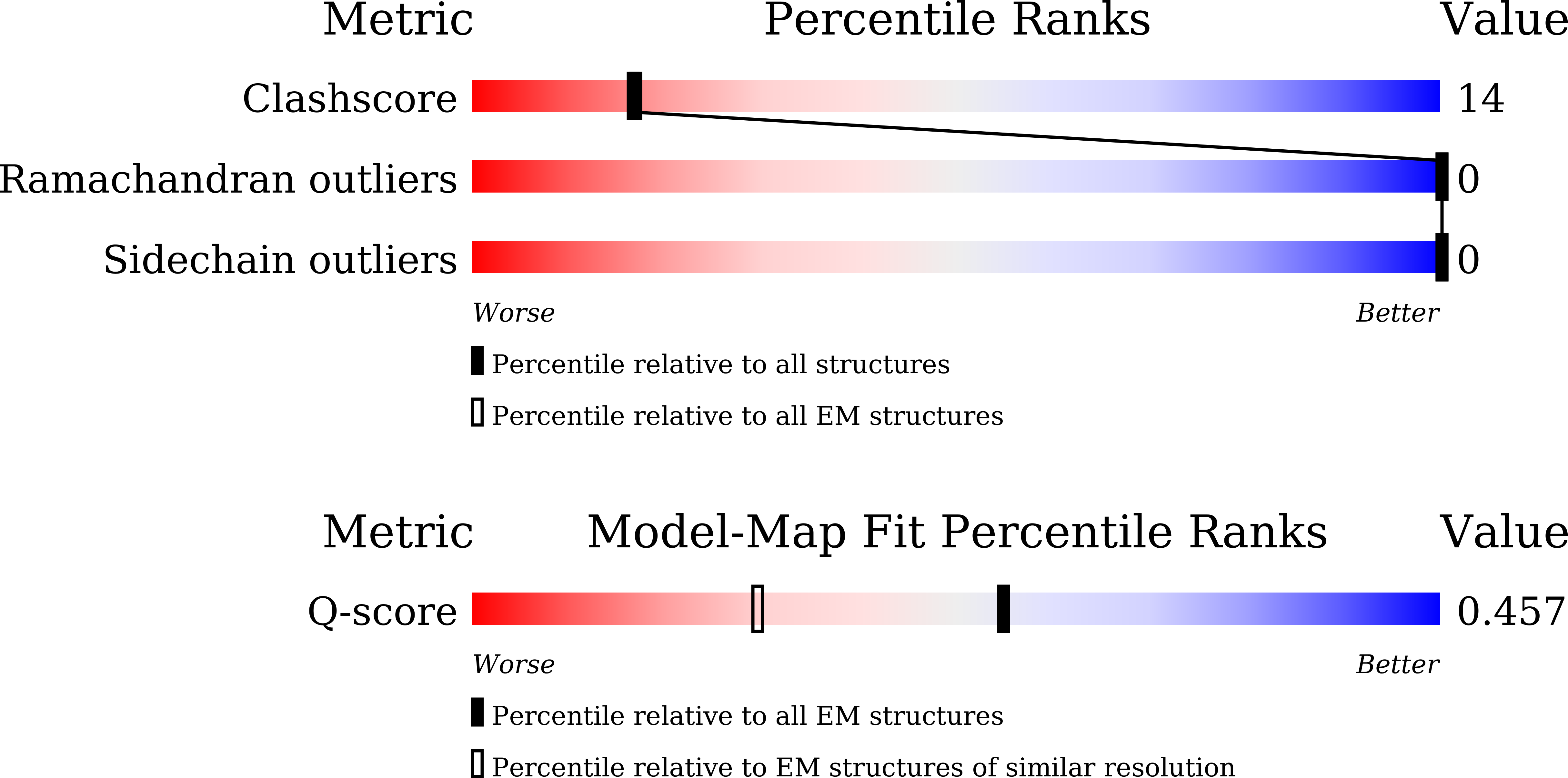
9K8S image
Deposition Date 2024-10-24
Release Date 2025-10-29
Last Version Date 2025-10-29
Entry Detail
PDB ID:
9K8S
Keywords:
Title:
Cryo-EM structure of HE30 polymorph 1
Biological Source:
Source Organism:
Homo sapiens (Taxon ID: 9606)
Method Details:
Experimental Method:
Resolution:
2.90 Å
Aggregation State:
FILAMENT
Reconstruction Method:
HELICAL
Macromolecular Entities
Structures with similar UniProt ID
Protein Blast
Polymer Type:polypeptide(L)
Molecule:Ninjurin-1
Gene (Uniprot):NINJ1
Chain IDs:A (auth: E), B (auth: F), C (auth: G), D (auth: H), E (auth: I), F (auth: J), G (auth: K), H (auth: L), I (auth: M), J (auth: N), K (auth: O), L (auth: P)
Chain Length:30
Number of Molecules:12
Biological Source:Homo sapiens
Ligand Molecules
Primary Citation
Amyloid Fibrillation of a Ninjurin-1-Derived alpha-Helical Peptide: Structural Insights into Conformational Transition.
Acs Nano 19 35977 35991 (2025)
PMID: 41037056 DOI: 10.1021/acsnano.5c14731

Abstact

Amyloid fibrils, defined by their cross-β architecture, are central to both disease and function, yet the molecular principles governing their formation remain incompletely understood. Ninjurin-1 (NINJ1), a membrane protein essential for plasma membrane rupture (PMR) during cell death, contains an N-terminal amphipathic α-helix. Here, we investigate a key peptide fragment of this region (residues 40-69, HE30) and uncover its membrane-disruptive activity, self-assembly, and structural transitions. Monomeric HE30 reorganizes lipids to induce membrane thinning while undergoing an environmentally responsive α-helix-to-β-sheet transition that drives amyloid fibril formation. Fibrils formed at physiological temperatures are predominantly nontwisted, but elevated temperatures induce left-handed twisted structures with variable pitches and lengths, and even result in high-order superhelical bundles. We further resolved the twisted fibril structures of HE30 by cryo-EM, revealing two distinct fibril polymorphs stabilized by both hydrophobic and electrostatic interactions. Consistently, salts inhibit HE30 fibrillation, emphasizing the role of electrostatic interactions in stabilizing fibrils. Moreover, acidic conditions (∼pH 4.4) promote fibril formation, whereas alkaline conditions lead to disassembly into α-helical monomers in a reversible manner. In situ AFM tracking reveals the asymmetric growth of fibrils, where one end elongates faster and the opposite end exhibits slower growth or complete inhibition. Functionally, HE30 fibrils are nontoxic and act as scaffolds for the temperature-controlled assembly of gold nanoparticle (AuNPs) superstructures. These findings not only advance our understanding of NINJ1-induced PMR but also provide a detailed structural basis for HE30 fibril formation via α-helix to β-sheet transitions and underscore their potential as building blocks for fibril-based biomaterials.

Legend

Protein

Chemical

Disease

Primary Citation of related structures