Deposition Date
2024-10-24
Release Date
2024-11-06
Last Version Date
2025-06-25
Entry Detail
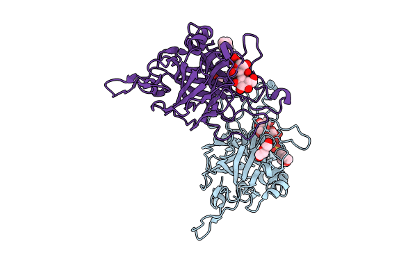
PDB ID:
9K7M
Keywords:
Title:
Coprinopsis cinerea GH131 protein CcGH131B E161A in complex with cellobiose
Biological Source:
Source Organism(s):
Expression System(s):
Method Details:
Experimental Method:
Resolution:
1.45 Å
R-Value Free:
0.18
R-Value Work:
0.15
R-Value Observed:
0.15
Space Group:
P 61