Deposition Date
2024-10-16
Release Date
2025-06-11
Last Version Date
2025-07-16
Entry Detail
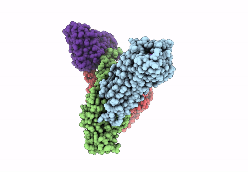
PDB ID:
9K20
Keywords:
Title:
Cryo-EM structure of ATP-bound P2Y purinoceptor 2-miniGo-scFv16 complex
Biological Source:
Source Organism(s):
Homo sapiens (Taxon ID: 9606)
Mus musculus (Taxon ID: 10090)
Mus musculus (Taxon ID: 10090)
Expression System(s):
Method Details:
Experimental Method:
Resolution:
2.65 Å
Aggregation State:
PARTICLE
Reconstruction Method:
SINGLE PARTICLE