9JZ1 image
Deposition Date 2024-10-13
Release Date 2025-10-15
Last Version Date 2025-11-05
Entry Detail
PDB ID:
9JZ1
Title:
Crystal structure of Nir2 C-terminal domain in complex with phosphate
Biological Source:
Source Organism:
Mus musculus (Taxon ID: 10090)
Host Organism:
Method Details:
Experimental Method:
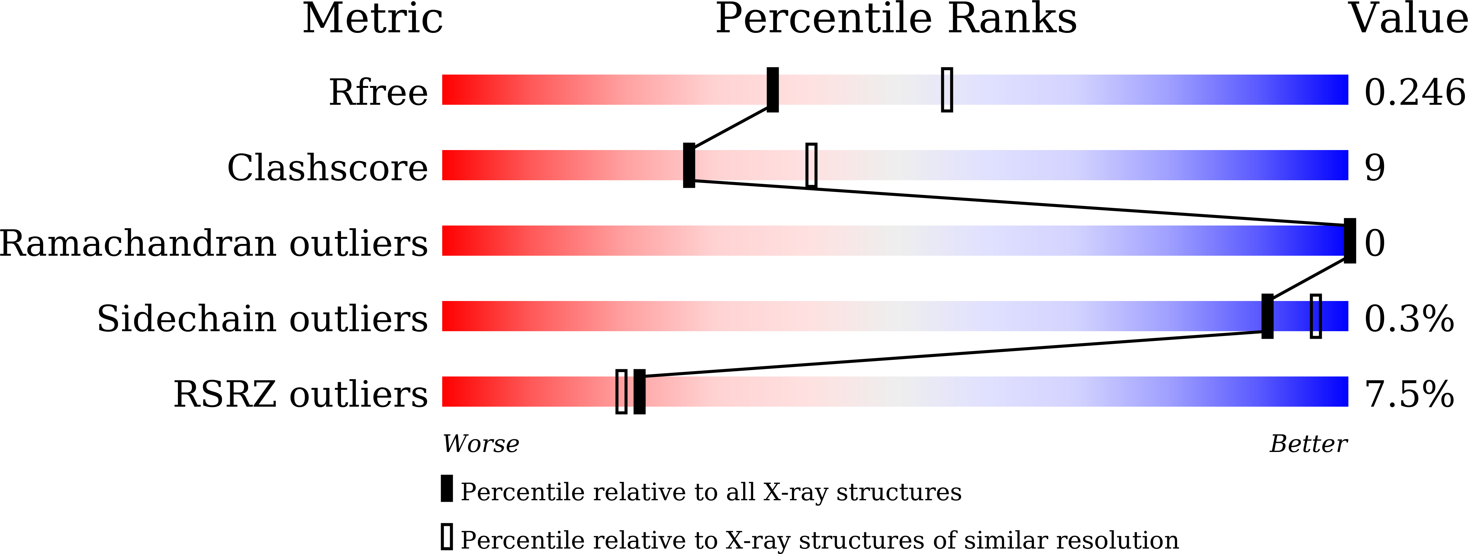
Resolution:
2.40 Å
R-Value Free:
0.26
R-Value Work:
0.21
R-Value Observed:
0.21
Space Group:
P 21 21 21
Macromolecular Entities
Structures with similar UniProt ID
Protein Blast
Polymer Type:polypeptide(L)
Molecule:Membrane-associated phosphatidylinositol transfer protein 1
Gene (Uniprot):Pitpnm1
Chain IDs:A, B, C
Chain Length:284
Number of Molecules:3
Biological Source:Mus musculus
Primary Citation
Nir2 crystal structures reveal a phosphatidic acid-sensing mechanism at ER-PM contact sites.
Proc.Natl.Acad.Sci.USA 122 e2516849122 e2516849122 (2025)
PMID: 41129229 DOI: 10.1073/pnas.2516849122

Abstact

Agonist-induced activation of phosphoinositide-specific phospholipase C (PLC) converts phosphatidylinositol 4,5-bisphosphate [PI(4,5)P2] to diacylglycerol (DAG) at the inner leaflet of the plasma membrane (PM). DAG can be enzymatically transformed into phosphatidic acid (PA) and accumulated at the PM. PYK2 N-terminal domain-interacting receptor 2 (Nir2) mediates the formation of ER-PM membrane contact sites (MCSs) by specifically recognizing PA at the PM and directly interacting with ER membrane protein vesicle-associated membrane protein-associated proteins (VAPs). The N-terminal phosphatidylinositol transfer protein domain of Nir2 facilitates PI/PA exchange at ER-PM MCSs to maintain PI and PA levels. Here, we reveal the mechanisms by which Nir2 senses phosphatidic acid (PA) and associates with membranes, based on three crystal structures of its C-terminal Lipin/Ned1/Smp2 (LNS2) domain bound to PA, the diphenylalanine [FF]-containing acidic tract (FFAT) motif complexed with vesicle-associated membrane protein-associated protein B/C (VAPB), and the Asp-Asp-His-Asp (DDHD) domain. The C-terminal LNS2 domain of Nir2 directly interacts with the phosphate in the headgroup of PA via hydrogen bonds involving S1025, T1065, K1103, and K1126. Formation of a salt bridge between E355 in Nir2 and R55 in VAPB is essential for Nir2 FFAT-VAPB interaction. The central DDHD domain of Nir2 forms a twofold symmetric dimer, and this self-association contributes to stable and tight membrane association. These findings reveal how Nir2-mediated ER-PM MCS formation maintains continued PI(4,5)P2-dependent PLC signaling.

Legend

Protein

Chemical

Disease

Primary Citation of related structures
Feedback Form
Name
Email
Institute
Feedback