9JVT image
Deposition Date 2024-10-09
Release Date 2025-10-15
Last Version Date 2025-12-17
Entry Detail
PDB ID:
9JVT
Title:
Structure of the C-terminal 4 domains (V4-V6-HP) villin bound to an actin
Biological Source:
Source Organism(s):
Expression System(s):
Method Details:
Experimental Method:
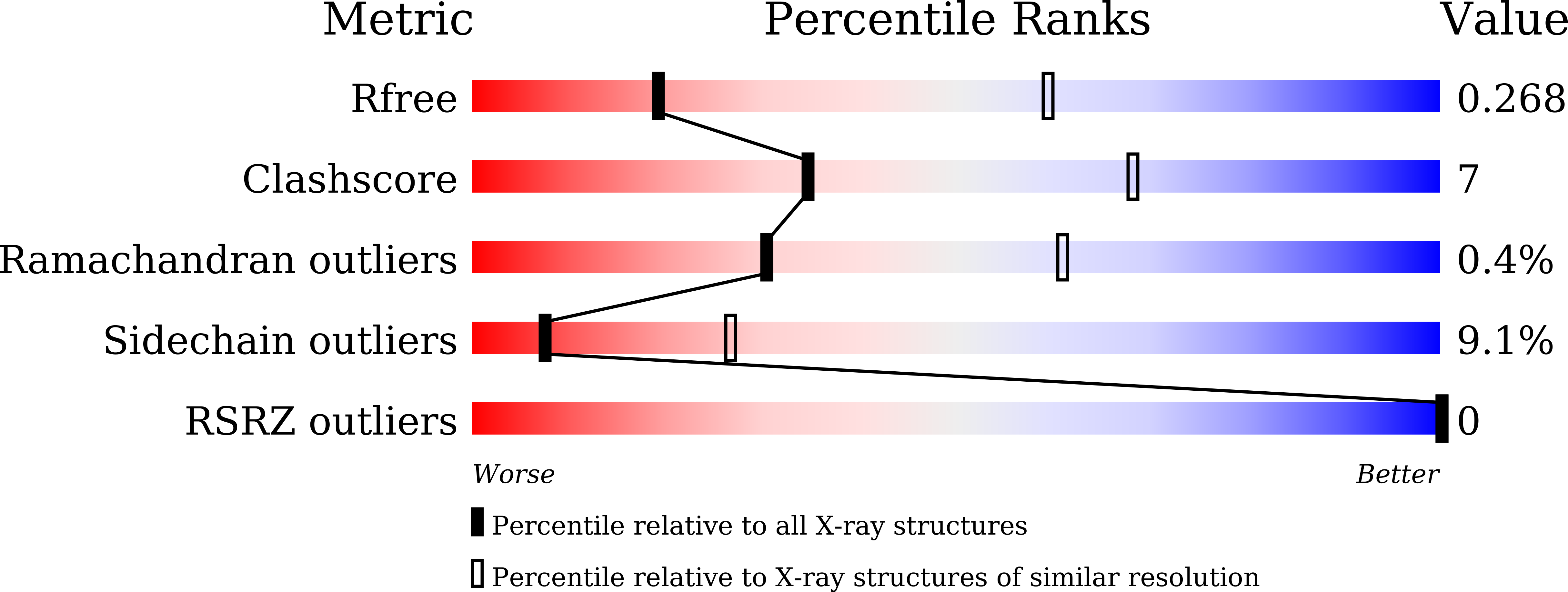
Resolution:
3.29 Å
R-Value Free:
0.26
R-Value Work:
0.24
R-Value Observed:
0.24
Space Group:
C 1 2 1
Macromolecular Entities
Structures with similar UniProt ID
Protein Blast
Polymer Type:polypeptide(L)
Molecule:Actin, alpha skeletal muscle
Gene (Uniprot):ACTA1
Chain IDs:B (auth: g), D (auth: G)
Chain Length:377
Number of Molecules:2
Biological Source:Gallus gallus
Polymer Type:polypeptide(L)
Molecule:Villin
Chain IDs:A (auth: v), C (auth: V)
Chain Length:485
Number of Molecules:2
Biological Source:Paralvinella sulfincola
Modified Residue
Compound ID Chain ID Parent Comp ID Details 2D Image
HIC B HIS modified residue
Primary Citation
The structure of an actin nucleus stabilized by villin.
Sci Adv 11 eadw6915 eadw6915 (2025)
PMID: 41337577 DOI: 10.1126/sciadv.adw6915

Abstact

Villin is an actin filament nucleating, severing, capping and bundling protein; however, the structural basis for villin's functions and the characteristics of the actin polymerization nucleus remain poorly understood. Here, we present the structure of vent-worm villin bound to a trimeric actin nucleus. Villin wraps around and caps the barbed end of the actin trimer. Its headpiece domain interacts at the junction of two laterally associated actin protomers, leaving the pointed-end subunits open for elongation. Within the actin trimer, the two longitudinally associated subunits adopt barbed and pointed-end subunit conformations, while the lateral protomer exhibits a monomeric conformation. This provides the first view of an actin-filament nucleus, revealing that the transition into the filamentous form is stimulated and stabilized by the interactions with the pointed-end subunits. Our results also illuminate mechanisms of actin-filament dynamics and villin capping and severing, suggesting that F-to-G actin conformational transitions facilitate the later process.

Legend

Protein

Chemical

Disease

Primary Citation of related structures
Feedback Form
Name
Email
Institute
Feedback