9JU0 image
Deposition Date 2024-10-07
Release Date 2025-09-03
Last Version Date 2025-09-03
Entry Detail
PDB ID:
9JU0
Keywords:
Title:
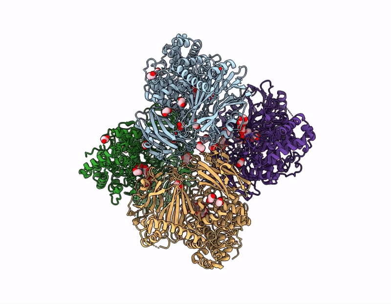
C-terminally truncated dextran dextrinase bound with acarbose
Biological Source:
Source Organism:
Host Organism:
Method Details:
Experimental Method:
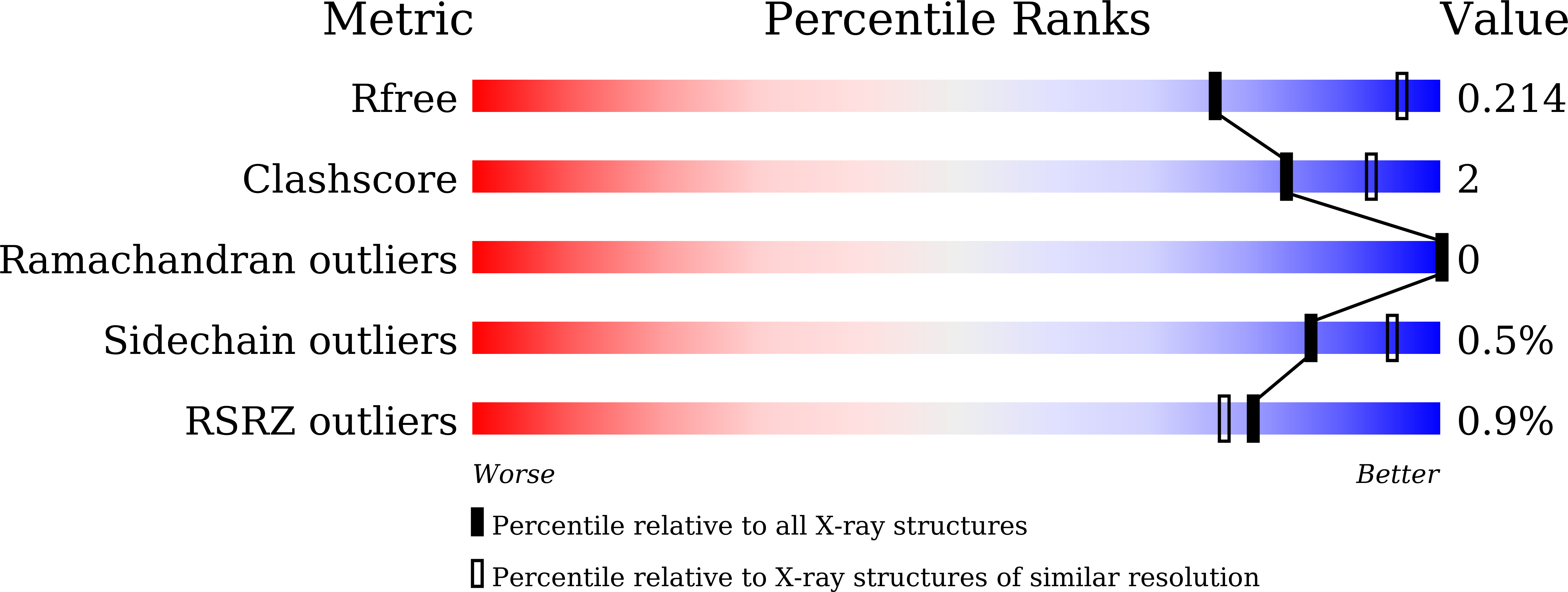
Resolution:
2.50 Å
R-Value Free:
0.21
R-Value Work:
0.18
Space Group:
P 21 21 21
Macromolecular Entities
Structures with similar UniProt ID
Protein Blast
Polymer Type:polypeptide(L)
Molecule:Dextran dextrinase
Gene (Uniprot):HKD20_12845
Chain IDs:A (auth: AAA), B (auth: BBB), C (auth: CCC), D (auth: DDD)
Chain Length:923
Number of Molecules:4
Biological Source:Gluconobacter oxydans
Peptide-like Molecules
PRD_900007
Primary Citation
Structural basis of transglucosylation in dextran dextrinase, a homolog of anomer-inverting GH15 glucoside hydrolases.
J.Biol.Chem. 301 110541 110541 (2025)
PMID: 40749827 DOI: 10.1016/j.jbc.2025.110541

Abstact

Bacterial exopolysaccharide, dextran, primarily composed of α-(1→6)-linked d-glucosyl residues, is synthesized from α-(1→4)-glucan dextrin or sucrose through successive anomer-retaining transglucosylation reactions by dextran dextrinase (DDase) or dextransucrase, respectively. Although the structure-function relationship of dextransucrase has been extensively studied, that of DDase remains largely unknown. Herein, we revealed the Gluconobacter oxydans DDase structural basis through biochemical and structural analyses. The DDase comprises 1284 residues, with its N-terminal 902 residues being functionally essential. Crystal structure analysis of the minimal active DDase (Δ382C) complex with the pseudo-maltotetraose inhibitor, acarbose, revealed its homodimeric structure. A Δ382C protomer contains two β-sandwich domains, N1 and N2, and an (α/α)6-barrel domain A. Surprisingly, domains N2, A, and the helix-loop-helix connecting them structurally resemble those of bacterial anomer-inverting glucohydrolases in glycoside hydrolase family 15 (GH15). Domain N1 primarily forms intra- and inter-subunit domain interfaces. The DDase acarbose-binding residues in subsite -1 are conserved with GH15 glucohydrolases. The DDase Glu671 and Glu858 are positioned similarly to the GH15 glucohydrolase general acid and base catalysts, respectively. However, Glu858 is approximately 1.2 to 1.6 Å closer to the acarbose equivalent anomeric carbon, facilitating its role as a nucleophilic catalyst in the double displacement mechanism. The catalytic residue functions were biochemically confirmed using mutant enzymes. Spatial position of Glu858 is arranged by the local structure of the α11→α12 loop and subunit interactions involving domain N1. Enzymes classified in the same GH family catalyze reactions with different mechanisms, anomer-inverting or -retaining, due to differences in their catalytic residue spatial arrangement.

Legend

Protein

Chemical

Disease

Primary Citation of related structures
Feedback Form
Name
Email
Institute
Feedback