9JRY image
Entry Detail
PDB ID:
9JRY
Title:
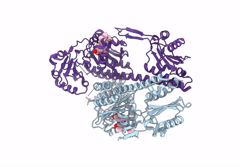
Crystal structure of FiDCB, a dual-cysteine cyanobacterial phytochrome of Fischerella sp. PCC 9605
Biological Source:
Source Organism:
Host Organism:
PDB Version:
Deposition Date:
2024-09-29
Release Date:
2025-01-22
Method Details:
Experimental Method:
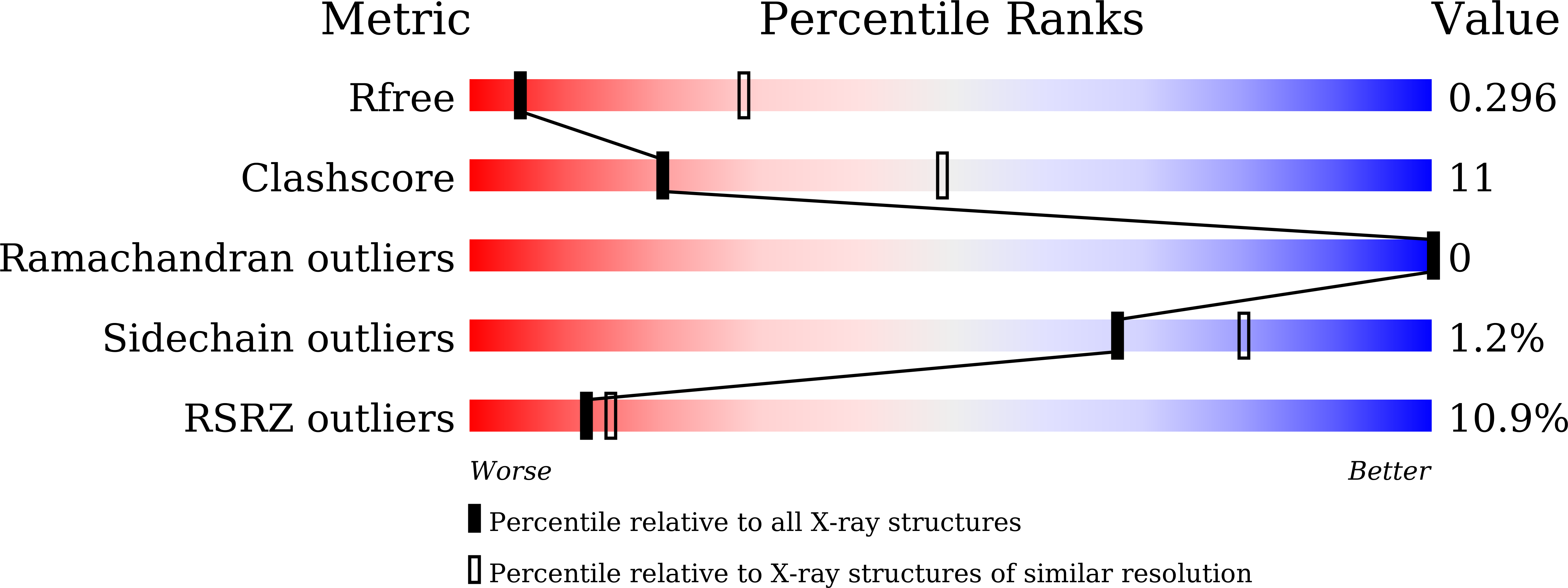
Resolution:
3.35 Å
R-Value Free:
0.29
R-Value Work:
0.27
R-Value Observed:
0.28
Space Group:
C 1 2 1
Macromolecular Entities
Polymer Type:polypeptide(L)
Description:FiDCB
Chain IDs:A, B
Chain Length:521
Number of Molecules:2
Biological Source:Fischerella sp. PCC 9605
Ligand Molecules
Primary Citation
Dual-Cys bacteriophytochromes: intermediates in cyanobacterial phytochrome evolution?
Febs J. 292 1197 1216 (2025)
PMID: 39801362 DOI: 10.1111/febs.17395

Abstact

Previous studies have identified three families of knotted phytochrome photoreceptors in cyanobacteria. We describe a fourth type: 'hybrid' phytochromes with putative bilin-binding cysteine residues in both their N-terminal 'knot' extensions and cGMP-phosphodiesterase/adenylate cyclase/FhlA (GAF) domains, which we designate as dual-cysteine bacteriophytochromes (DCBs). Recombinant expression of DCBs in Escherichia coli yields photoactive phycocyanobilin (PCB) adducts with red/far-red photocycles similar to those of the GAF-Cys-containing cyanobacterial phytochromes (Cph1s). Incorporation of the PCB precursor, biliverdin IXα (BV), gave multiple populations, one of which appears similar to those of cyanobacterial bacteriophytochromes (cBphPs). A crystal structure of FiDCB bound to BV exhibits two thioether linkages between the GAF- and 'PAS-knot'-Cys residues and the C31 and C32 atoms of BV. When expressed in Synechocystis sp. PCC 6803, DCBs incorporate PCB rather than BV. DCBs can be converted to photoactive cBphP-, Cph1-, and tandem-cysteine cyanobacterial phytochrome (TCCP) analogs by removal and/or addition of a cysteine residue by site-directed mutagenesis. This structural plasticity contrasts with our inability to generate functional photosensor analogs by analogous site-directed mutagenesis of TCCP and Cph1 representatives. Phylogenetic analysis demonstrates that DCBs do not form a monophyletic clade and also suggest that Cph1 and TCCP families independently emerged from different lineages of cBphPs, possibly via DCB intermediates.

Legend

Protein

Chemical

Disease

Primary Citation of related structures