9JFK image
Deposition Date 2024-09-04
Release Date 2025-12-03
Last Version Date 2025-12-03
Entry Detail
PDB ID:
9JFK
Title:
Cryo-EM structure of [Pen5]-urotensin (4-11)-bounded human Urotensin receptor (UTS2R)-Gq complex
Biological Source:
Source Organism:
Homo sapiens (Taxon ID: 9606)
Lama glama (Taxon ID: 9844)
synthetic construct (Taxon ID: 32630)
Host Organism:
Method Details:
Experimental Method:
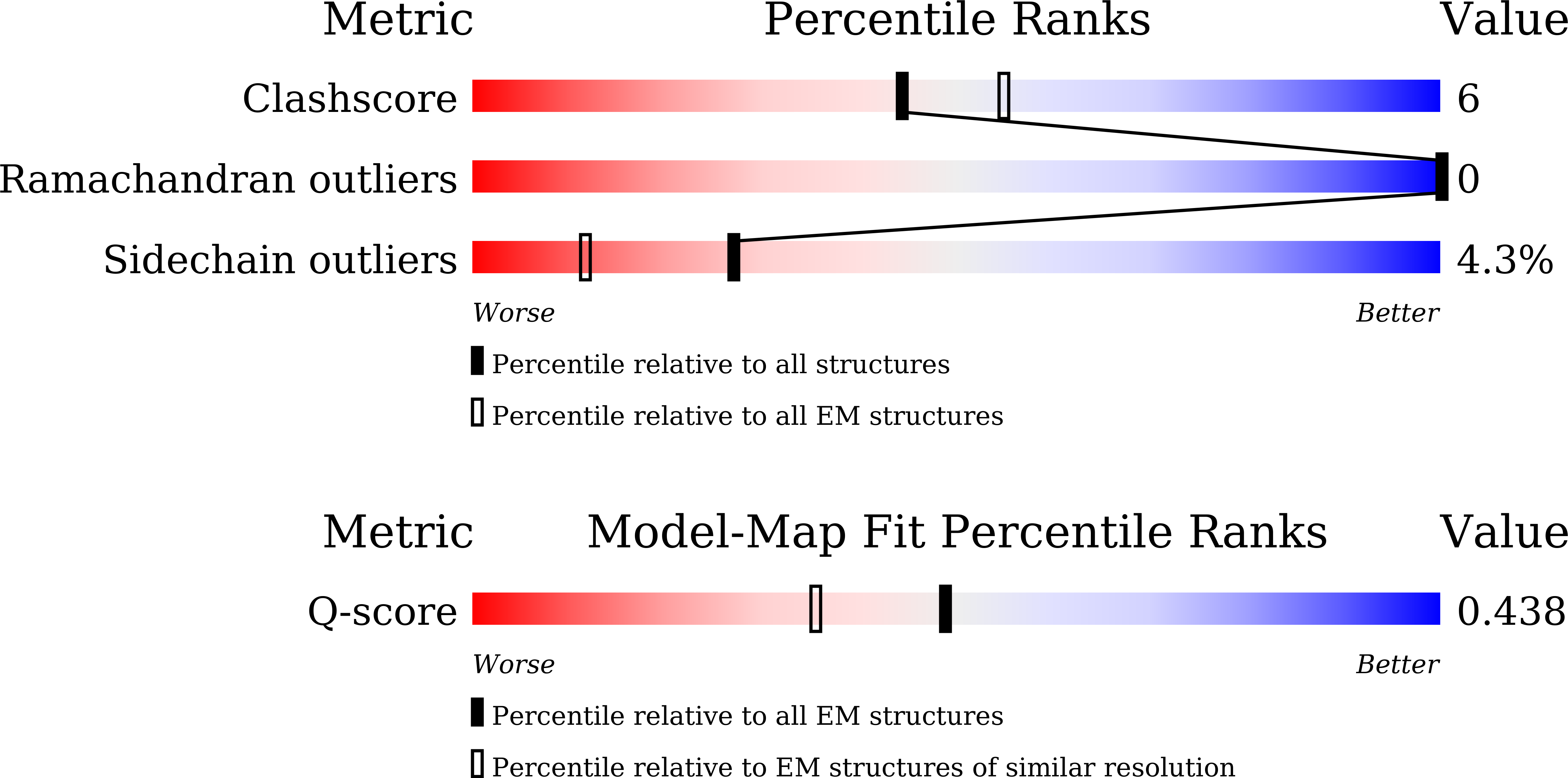
Resolution:
3.23 Å
Aggregation State:
PARTICLE
Reconstruction Method:
SINGLE PARTICLE
Macromolecular Entities
Structures with similar UniProt ID
Protein Blast
Polymer Type:polypeptide(L)
Molecule:Guanine nucleotide-binding protein G(I)/G(S)/G(T) subunit beta-1
Gene (Uniprot):GNB1
Chain IDs:A
Chain Length:345
Number of Molecules:1
Biological Source:Homo sapiens
Polymer Type:polypeptide(L)
Molecule:G subunit q (gi2-mini-gq chimeric)
Chain IDs:B
Chain Length:246
Number of Molecules:1
Biological Source:Homo sapiens
Polymer Type:polypeptide(L)
Molecule:ASP-LE1-PHE-TRP-LYS-TYR-CYS-VAL
Chain IDs:F (auth: C)
Chain Length:8
Number of Molecules:1
Biological Source:synthetic construct
Structures with similar UniProt ID
Protein Blast
Polymer Type:polypeptide(L)
Molecule:Guanine nucleotide-binding protein G(I)/G(S)/G(O) subunit gamma-2
Gene (Uniprot):GNG2
Chain IDs:C (auth: D)
Chain Length:70
Number of Molecules:1
Biological Source:Homo sapiens
Polymer Type:polypeptide(L)
Molecule:Nano body 35
Chain IDs:D (auth: N)
Chain Length:128
Number of Molecules:1
Biological Source:Lama glama
Structures with similar UniProt ID
Protein Blast
Polymer Type:polypeptide(L)
Molecule:Urotensin-2 receptor
Gene (Uniprot):UTS2R
Chain IDs:E (auth: R)
Chain Length:347
Number of Molecules:1
Biological Source:Homo sapiens
Ligand Molecules
Primary Citation
Structural insights into hormone recognition and G-protein coupling of the urotensin-II receptor.
J.Biol.Chem. 301 110794 110794 (2025)
PMID: 41062066 DOI: 10.1016/j.jbc.2025.110794

Abstact

Urotensin-II (U-II) is a potent vasoconstrictor peptide that interacts with the human urotensin-II receptor (UTR), a class A G protein-coupled receptor (GPCR) that primarily couples with Gq proteins. In this study, we present the cryo-electron microscopy structure of the miniGq-coupled UTR bound to the potent UTR agonist P5U, providing insights into unique ligand recognition and activation mechanisms. Unlike typical linear peptides, the cyclic structure of P5U engages the receptor's transmembrane domains through key side chain interactions involving residues F6, W7, K8, and Y9, which are crucial for receptor activation. Comparative analysis with somatostatin receptors (SSTRs) reveals distinct ligand specificity, driven by variations in side chain composition. Notably, we identify F2746.51 as the toggle switch residue in UTR, in contrast to the classical W6.48 seen in other GPCRs. Our findings elucidate the structural basis for UTR's Gq coupling specificity, highlighting unique Gαq interactions. This study advances the understanding of U-II signaling and offers a foundation for developing selective UTR modulators, with potential therapeutic implications for cardiovascular diseases linked to dysregulated U-II activity.

Legend

Protein

Chemical

Disease

Primary Citation of related structures
Feedback Form
Name
Email
Institute
Feedback