9JEW image
Entry Detail
PDB ID:
9JEW
Title:
Crystal structure of a cupin protein (tm1459, C106V mutant) in iron (Fe) substituted form
Biological Source:
Source Organism:
PDB Version:
Deposition Date:
2024-09-03
Release Date:
2025-05-07
Method Details:
Experimental Method:
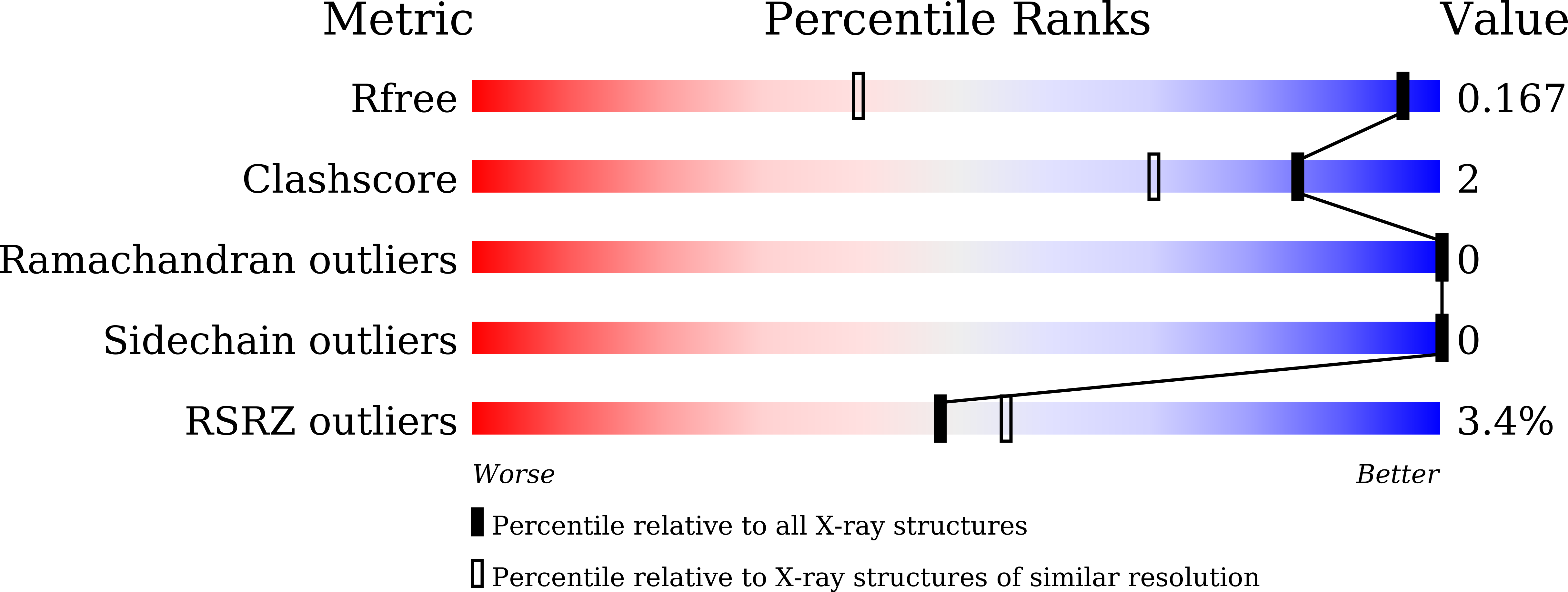
Resolution:
1.08 Å
R-Value Free:
0.16
R-Value Work:
0.13
R-Value Observed:
0.13
Space Group:
P 21 21 21
Macromolecular Entities
Polymer Type:polypeptide(L)
Description:Cupin type-2 domain-containing protein
Mutations:C106V
Chain IDs:A, B
Chain Length:118
Number of Molecules:2
Biological Source:Thermotoga maritima
Ligand Molecules
Primary Citation
Unusual Self-Hydroxylation in 4-Histidine Tetrad-Supporting Nonheme Iron Center.
Chem Asian J ? e202401191 e202401191 (2025)
PMID: 40260495 DOI: 10.1002/asia.202401191

Abstact

The TM1459 protein from Thermotoga maritima is a member of the cupin protein superfamily and contains a mononuclear metal center. Structural information has been obtained using X-ray crystallography; however, its physiological role remains unknown. The metal-binding site has an octahedral coordination geometry ligated by four histidine imidazoles and two terminal water molecules present in the cis position. This protein had the ability to bind Mn, Fe, and Zn ions; additionally, a self-hydroxylation reaction occurred in the Fe-TM1459 C106V mutant. Namely, one of the tyrosine residues (Tyr7) was hydroxylated to generate the green form. Spectroscopic analyses using Vis-NIR, MALDI-TOF/MS, and resonance Raman spectroscopy confirmed that Tyr7 was hydroxylated to 3,4-dihydroxyphenylalanine giving an iron-catecholate complex. Because the Y7A/C106V mutant did not produce this green form, the mutation of Cys106 to Val was assumed to have induced a conformational change in Tyr7 that facilitated its approach to the metal center promoting the self-hydroxylation reaction. Thus, these results demonstrated that Fe-TM1459 protein has monooxygenase activity.

Legend

Protein

Chemical

Disease

Primary Citation of related structures