9JBD image
Deposition Date 2024-08-26
Release Date 2025-08-27
Last Version Date 2025-08-27
Entry Detail
PDB ID:
9JBD
Keywords:
Title:
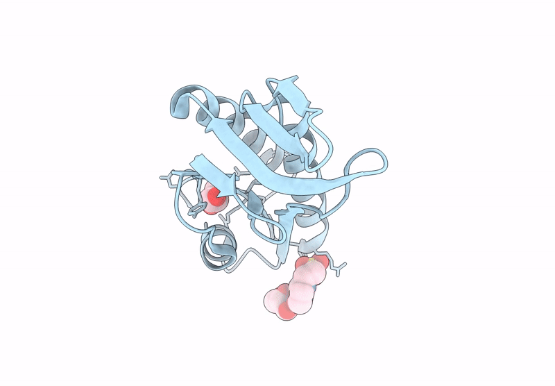
Crystal Structure of the MoaE-like domain within Rv3323c from Mycobacterium tuberculosis
Biological Source:
Method Details:
Experimental Method:
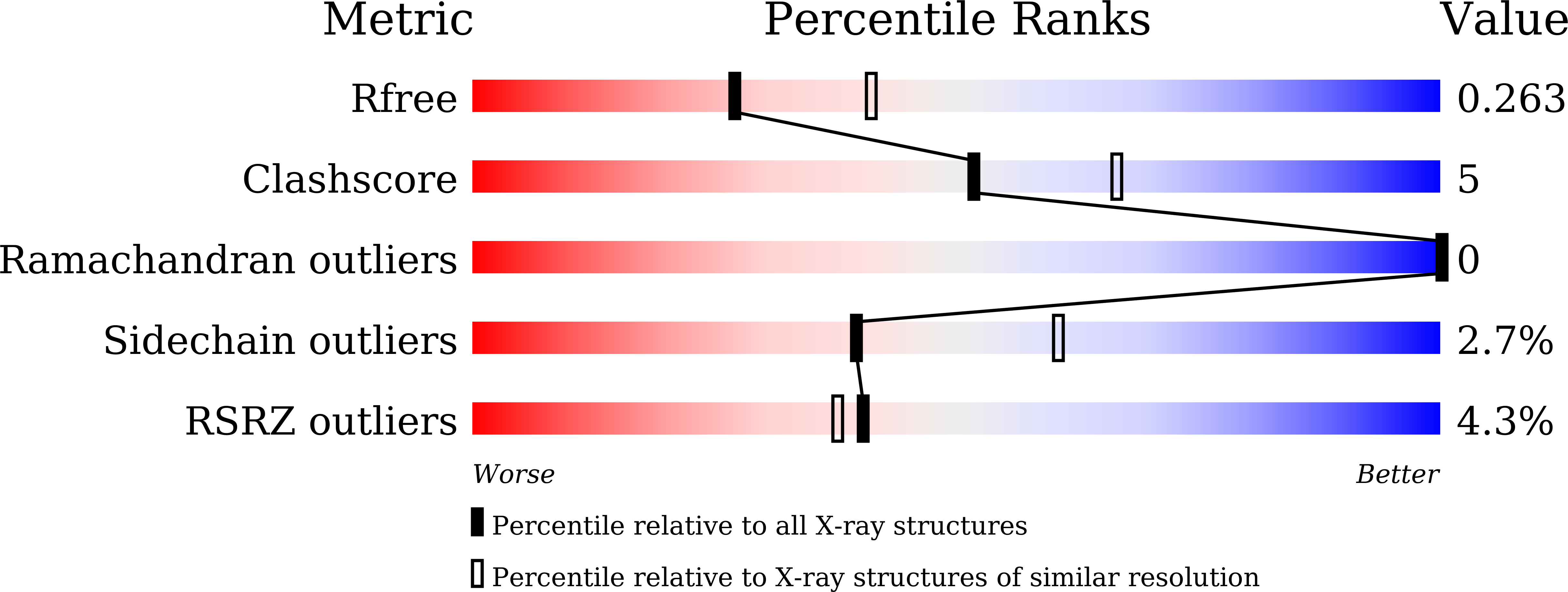
Resolution:
2.40 Å
R-Value Free:
0.26
R-Value Work:
0.21
R-Value Observed:
0.22
Space Group:
P 65 2 2
Macromolecular Entities
Structures with similar UniProt ID
Protein Blast
Polymer Type:polypeptide(L)
Molecule:Probable MoaD-MoaE fusion protein MoaX
Gene (Uniprot):moaX
Chain IDs:A
Chain Length:146
Number of Molecules:1
Biological Source:Mycobacterium tuberculosis (strain ATCC 25618 / H37Rv)
Primary Citation
Structural comparison of three MoaE proteins in Mycobacterium tuberculosis: Insights into molybdopterin synthase assembly and specificity.
Biochem.Biophys.Res.Commun. 768 151945 151945 (2025)
PMID: 40345009 DOI: 10.1016/j.bbrc.2025.151945

Abstact

Molybdoenzymes are essential for the survival and pathogenicity of Mycobacterium tuberculosis and require the molybdenum cofactor (MoCo). The biosynthesis of MoCo involves the molybdopterin (MPT) synthase complex, which is composed of the MoaD and MoaE subunits. The genome of M. tuberculosis encodes three homologs of MoaE: MoaE1, MoaE2, and MoaXE (the latter being a MoaE component of a MoaD-MoaE fusion protein known as MoaX), as well as three MoaD proteins. However, the structural basis for their functional specificity and interaction with MoaD partners remains unclear. We determined the crystal structures of all three MoaE proteins, revealing a conserved α/β hammerhead fold with distinct binding interface features resulting from minor sequence variations. Pull-down assays demonstrate that MoaE2 and MoaXE selectively interact with their cognate MoaD partners, while MoaE1 exhibits promiscuous binding to all MoaD forms. Although the structural plasticity of MoaE1 enables binding to three MoaD forms, it suggests that not all MoaE-MoaD combinations yield functional MPT synthase complexes, as structural rearrangements can lead to enzymatic inactivation. Our findings provide detailed insights into the molecular determinants that govern the assembly and specificity of MPT synthase in M. tuberculosis.

Legend

Protein

Chemical

Disease

Primary Citation of related structures
Feedback Form
Name
Email
Institute
Feedback