9J8K image
Deposition Date 2024-08-21
Release Date 2025-07-02
Last Version Date 2025-07-02
Entry Detail
PDB ID:
9J8K
Title:
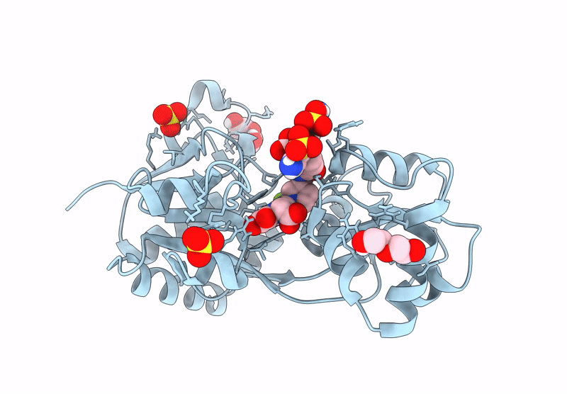
Crystal structure of the GluA2 ligand binding core (S1S2J) in complex with fluorophore-ligand conjugate
Biological Source:
Source Organism:
Host Organism:
Method Details:
Experimental Method:
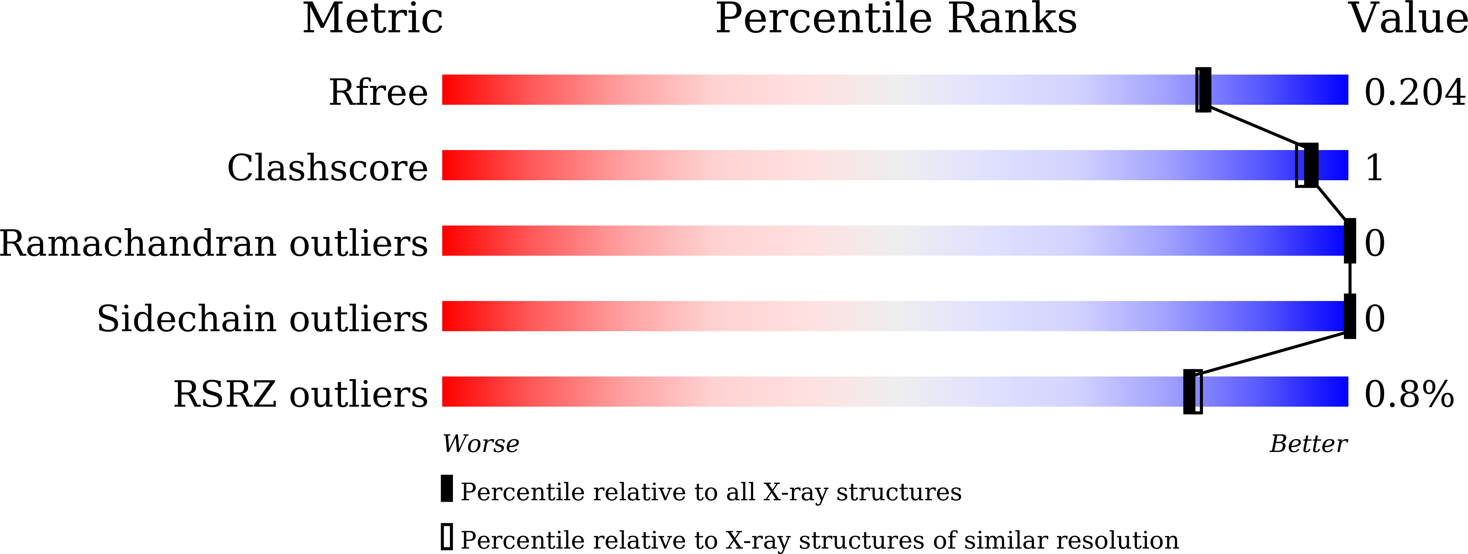
Resolution:
1.82 Å
R-Value Free:
0.19
R-Value Work:
0.16
R-Value Observed:
0.17
Space Group:
P 61 2 2
Macromolecular Entities
Polymer Type:polypeptide(L)
Molecule:GluA2 ligand binding core
Chain IDs:A
Chain Length:272
Number of Molecules:1
Biological Source:Rattus norvegicus
Primary Citation
Rapid and reversible fluorescent probe enables repeated snapshot imaging of AMPA receptors during synaptic plasticity.
Sci Adv 11 eadt6683 eadt6683 (2025)
PMID: 40479050 DOI: 10.1126/sciadv.adt6683

Abstact

The subcellular localization of neurotransmitter receptors is strictly regulated in neurons. Changes in the trafficking of α-amino-3-hydroxy-5-methyl-4-isoxazolepropionic acid (AMPA)-type glutamate receptors (AMPARs) play an essential role in synaptic plasticity, which is the cellular basis of learning and memory. To explore receptor trafficking, genetically encoded approaches (e.g., the fusion of fluorescent proteins to receptors) are often used. However, concerns remain that genetic approaches cannot fully reproduce the receptor functions that are inherent to neurons. Herein, we report on PFQX1(AF488), a fluorescent probe for the visualization of cell-surface AMPARs without any genetic manipulation to neurons. The rapid and reversible staining features of this probe enabled snapshot imaging, which showed the accumulation of native AMPARs in dendritic spines during synaptic plasticity. Moreover, the mechanism of this synaptic accumulation, for which genetically encoded approaches have given controversial results, was revealed by integrating two chemical methods: PFQX1(AF488) and covalent chemical labeling.

Legend

Protein

Chemical

Disease

Primary Citation of related structures