9IVA image
Entry Detail
PDB ID:
9IVA
EMDB ID:
Keywords:
Title:
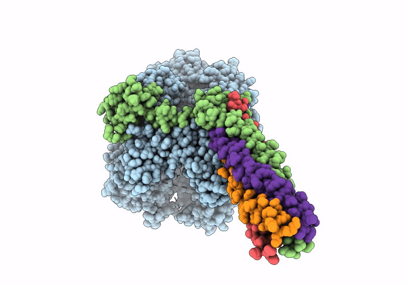
Cryo-EM structure of the full-length Nipah Virus L Protein bound by Phosphoprotein Tetramer
Biological Source:
Source Organism:
Host Organism:
PDB Version:
Deposition Date:
2024-07-23
Release Date:
2025-05-21
Method Details:
Experimental Method:
Resolution:
2.52 Å
Aggregation State:
PARTICLE
Reconstruction Method:
SINGLE PARTICLE
Macromolecular Entities
Polymer Type:polypeptide(L)
Description:RNA-directed RNA polymerase L
Chain IDs:A
Chain Length:2244
Number of Molecules:1
Biological Source:Henipavirus nipahense
Polymer Type:polypeptide(L)
Description:Phosphoprotein
Chain IDs:B, C, D, E
Chain Length:709
Number of Molecules:4
Biological Source:Henipavirus nipahense
Ligand Molecules
Primary Citation
Cryo-EM structures of Nipah virus polymerase complex reveal highly varied interactions between L and P proteins among paramyxoviruses.
Protein Cell ? ? ? (2025)
PMID: 39964914 DOI: 10.1093/procel/pwaf014

Abstact

Nipah virus (NiV) and related viruses form a distinct henipavirus genus within the Paramyxoviridae family. NiV continues to spillover into the humans causing deadly outbreaks with increasing human-bat interaction. NiV encodes the large protein (L) and phosphoprotein (P) to form the viral RNA polymerase machinery. Their sequences show limited homologies to those of non-henipavirus paramyxoviruses. We report two cryo-electron microscopy (cryo-EM) structures of the Nipah virus (NiV) polymerase L-P complex, expressed and purified in either its full-length or truncated form. The structures resolve the RNA-dependent RNA polymerase (RdRp) and polyribonucleotidyl transferase (PRNTase) domains of the L protein, as well as a tetrameric P protein bundle bound to the L-RdRp. L-protein C-terminal regions are unresolved, indicating flexibility. Two PRNTase domain zinc-binding sites, conserved in most Mononegavirales, are confirmed essential for NiV polymerase activity. The structures further reveal anchoring of the P protein bundle and P protein X domain (XD) linkers on L, via an interaction pattern distinct among Paramyxoviridae. These interactions facilitate binding of a P protein XD linker in the nucleotide entry channel and distinct positioning of other XD linkers. We show that the disruption of the L-P interactions reduces NiV polymerase activity. The reported structures should facilitate rational antiviral-drug discovery and provide a guide for the functional study of NiV polymerase.

Legend

Protein

Chemical

Disease

Primary Citation of related structures