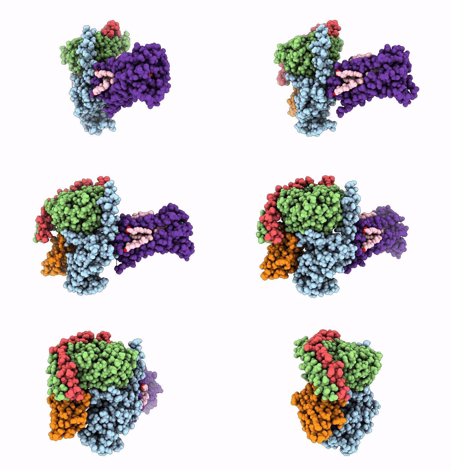
9ITB image
Deposition Date 2024-07-19
Release Date 2025-01-15
Last Version Date 2025-02-05
Entry Detail
PDB ID:
9ITB
Title:
LPA-bound LPAR6 in complex with miniGq
Biological Source:
Source Organism:
Homo sapiens (Taxon ID: 9606)
Lama glama (Taxon ID: 9844)
Host Organism:
Method Details:
Experimental Method:
Resolution:
2.89 Å
Aggregation State:
PARTICLE
Reconstruction Method:
SINGLE PARTICLE
Macromolecular Entities
Polymer Type:polypeptide(L)
Molecule:engineered miniGaq
Chain IDs:A
Chain Length:362
Number of Molecules:1
Biological Source:Homo sapiens
Polymer Type:polypeptide(L)
Molecule:Guanine nucleotide-binding protein G(I)/G(S)/G(T) subunit beta-1
Gene (Uniprot):GNB1
Chain IDs:B
Chain Length:345
Number of Molecules:1
Biological Source:Homo sapiens
Polymer Type:polypeptide(L)
Molecule:Guanine nucleotide-binding protein G(I)/G(S)/G(O) subunit gamma-2
Gene (Uniprot):GNG2
Chain IDs:E (auth: G)
Chain Length:71
Number of Molecules:1
Biological Source:Homo sapiens
Polymer Type:polypeptide(L)
Molecule:Nb35
Chain IDs:C (auth: N)
Chain Length:161
Number of Molecules:1
Biological Source:Lama glama
Polymer Type:polypeptide(L)
Molecule:Lysophosphatidic acid receptor 6
Gene (Uniprot):LPAR6
Chain IDs:D (auth: R)
Chain Length:294
Number of Molecules:1
Biological Source:Homo sapiens
Primary Citation
Molecular mechanism of ligand recognition and activation of lysophosphatidic acid receptor LPAR6.
Proc.Natl.Acad.Sci.USA 122 e2415426122 e2415426122 (2025)
PMID: 39847322 DOI: 10.1073/pnas.2415426122

Abstact

Lysophosphatidic acid (LPA) exerts its physiological roles through the endothelialdifferentiation gene (EDG) family LPA receptors (LPAR1-3) or the non-EDG family LPA receptors (LPAR4-6). LPAR6 plays crucial roles in hair loss and cancer progression, yet its structural information is very limited. Here, we report the cryoelectron microscopy structure of LPA-bound human LPAR6 in complex with a mini G13 or Gq protein. These structures reveal a distinct ligand binding and recognition mode that differs significantly from that of LPAR1. Specifically, LPA uses its charged head to form an extensive polar interaction network with key polar residues on the extracellular side of transmembrane helix 5-6 and the extracellular loop 2. Structural comparisons and homology analysis suggest that the EDG and non-EDG families use two distinct modes for LPA binding. The structural observations are validated through functional mutagenesis studies. We further uncover the mechanisms of LPAR6 activation and principles of G-protein coupling. The structural information revealed by our study lays the groundwork for understanding LPAR6 signaling and provides a rational basis for designing compounds targeting LPAR6.

Legend

Protein

Chemical

Disease

Primary Citation of related structures