9IR6 image
Entry Detail
PDB ID:
9IR6
Keywords:
Title:
Crystal structure of UDP-N-acetylmuramic Acid L-alanine ligase (MurC) from Roseburia faecis in complex with UNAM
Biological Source:
Source Organism:
PDB Version:
Deposition Date:
2024-07-14
Release Date:
2025-04-09
Method Details:
Experimental Method:
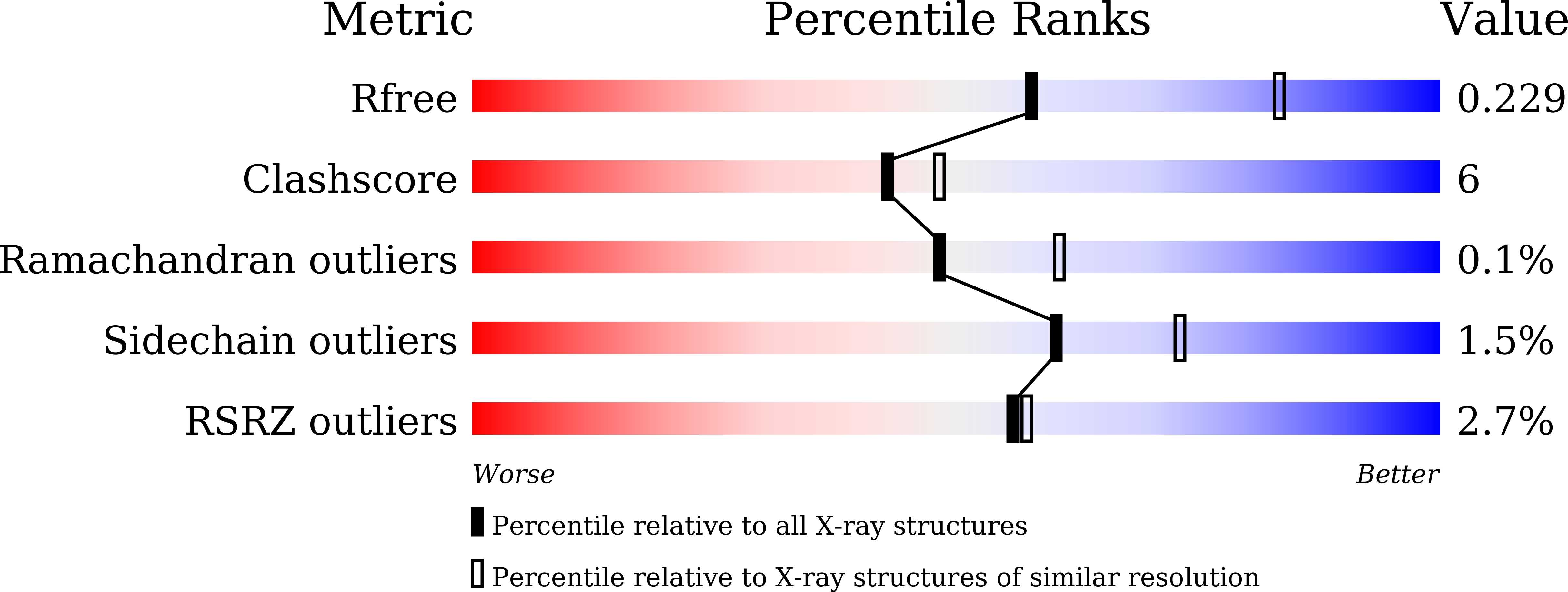
Resolution:
2.43 Å
R-Value Free:
0.22
R-Value Work:
0.18
R-Value Observed:
0.19
Space Group:
P 1 21 1
Macromolecular Entities
Polymer Type:polypeptide(L)
Description:UDP-N-acetylmuramate--L-alanine ligase
Chain IDs:A, B
Chain Length:460
Number of Molecules:2
Biological Source:Roseburia faecis
Primary Citation
Unusual MurC Ligase and Peptidoglycan Discovered in Lachnospiraceae Using a Fluorescent L-Amino Acid Based Selective Labeling Probe.
Angew.Chem.Int.Ed.Engl. ? e202503049 e202503049 (2025)
PMID: 40152026 DOI: 10.1002/anie.202503049

Abstact

Developing selective labeling probes for specific bacterial taxa can not only facilitate the study of target bacteria but also deepen our understanding of the microbial diversity at structural and molecular levels. The availability of such probes, however, remains very limited. In this study, by exploiting the variation of amino acids in peptidoglycan stem peptide, we designed a fluorescent L-amino acid probe and found that it can selectively target the family Lachnospiraceae (a major Gram-positive family in murine gut microbiome) in vivo. The following in vitro test using two Roseburia species belonging to this family validated labeling by the probe. We then discovered that the labeling site is the first amino acid (L-alanine in most bacteria), which links the stem peptide with N-acetylmuramic acid, a process catalyzed by a highly conserved enzyme MurC. An enzyme assay of Roseburia MurC demonstrated its ability to conjugate a fluorescent L-amino acid and other non-L-Ala amino acids to UDP-N-acetylmuramic acid. Subsequent X-ray crystallography analysis uncovered a substantially enlarged inner space in this enzyme, which can partially explain its tolerance to these atypical substrates. The resulting unusual peptidoglycan structures lead to significantly reduced activation of the NOD immune receptors, suggesting a new mechanism for the host to accommodate these highly abundant commensals.

Legend

Protein

Chemical

Disease

Primary Citation of related structures