9IOQ image
Entry Detail
PDB ID:
9IOQ
Title:
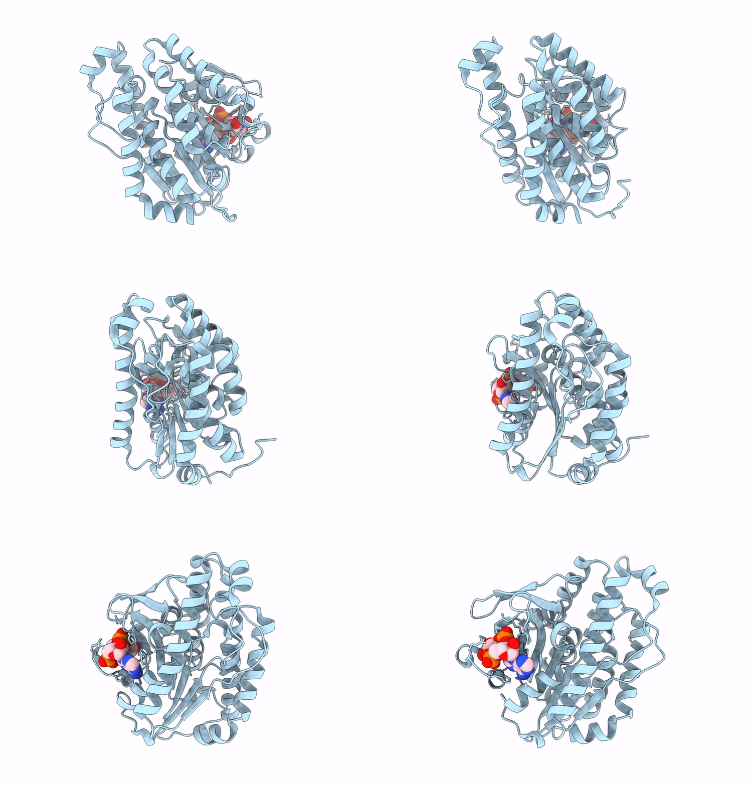
Crystal structure of CapE bound cUA
Biological Source:
Source Organism:
Host Organism:
PDB Version:
Deposition Date:
2024-07-09
Release Date:
2025-05-21
Method Details:
Experimental Method:
Resolution:
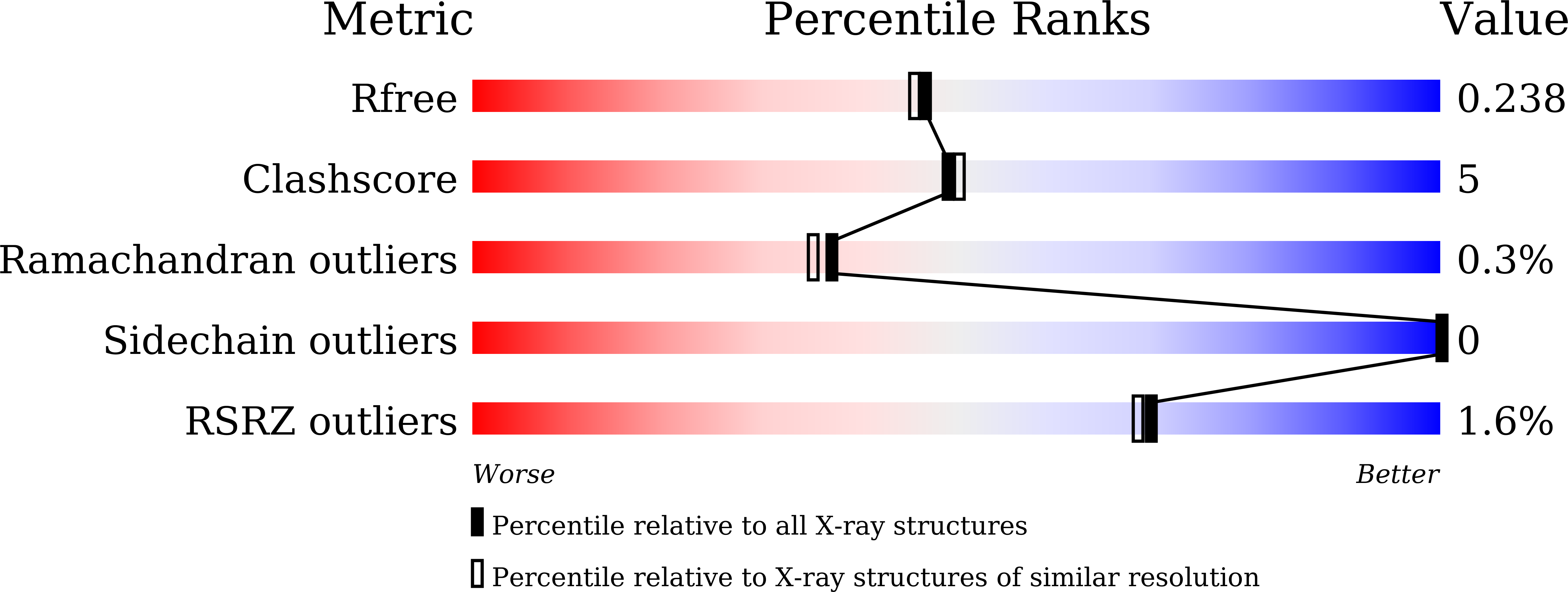
2.00 Å
R-Value Free:
0.23
R-Value Work:
0.19
R-Value Observed:
0.19
Space Group:
P 32 2 1
Macromolecular Entities
Polymer Type:polypeptide(L)
Description:cUMP-AMP-activated phospholipase
Chain IDs:A
Chain Length:320
Number of Molecules:1
Biological Source:Escherichia coli
Ligand Molecules
Primary Citation
Cyclic-dinucleotide-induced filamentous assembly of phospholipases governs broad CBASS immunity.
Cell ? ? ? (2025)
PMID: 40345202 DOI: 10.1016/j.cell.2025.04.022

Abstact

Cyclic-oligonucleotide-based antiphage signaling systems (CBASS), a widespread antiviral bacterial immune system homologous to the mammalian cGAS-STING pathway, synthesizes cyclic nucleotide signals and triggers effector proteins to induce cell death and prevent viral propagation. Among various CBASS effectors, phospholipase effectors are the first to be discovered and are one of the most widespread families that sense cyclic dinucleotides to degrade cell membrane phospholipids. Here, we report that CBASS phospholipases assemble from a dimeric inactive state into active higher-order filamentous oligomers upon sensing cyclic dinucleotides. Using a combined approach of cryo-electron microscopy and X-ray crystallography, we have determined the structures of CBASS phospholipase in the inactive dimeric state, the cyclic-dinucleotide-bound active higher-order state, and the substrate-analog-bound catalytic mimicry state, thereby visualizing the complete conformational reorganization process. Complemented by functional assays of intermolecular binding, phospholipase enzymatic activity, in vitro membrane disruption, and in vivo antiphage efficiency, our work elucidates the mechanisms of assembly and activation of CBASS phospholipases.

Legend

Protein

Chemical

Disease

Primary Citation of related structures