Deposition Date
2024-07-01
Release Date
2025-03-19
Last Version Date
2025-03-19
Entry Detail
PDB ID:
9ILT
Keywords:
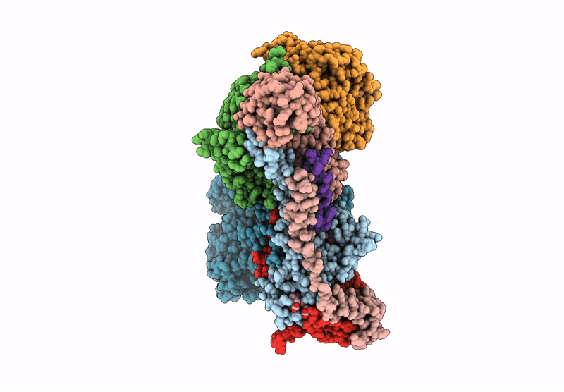
Title:
Crystal structure of alternative complex III from Chloroflexus aurantiacus
Biological Source:
Source Organism(s):
Chloroflexus aurantiacus J-10-fl (Taxon ID: 324602)
Method Details:
Experimental Method:
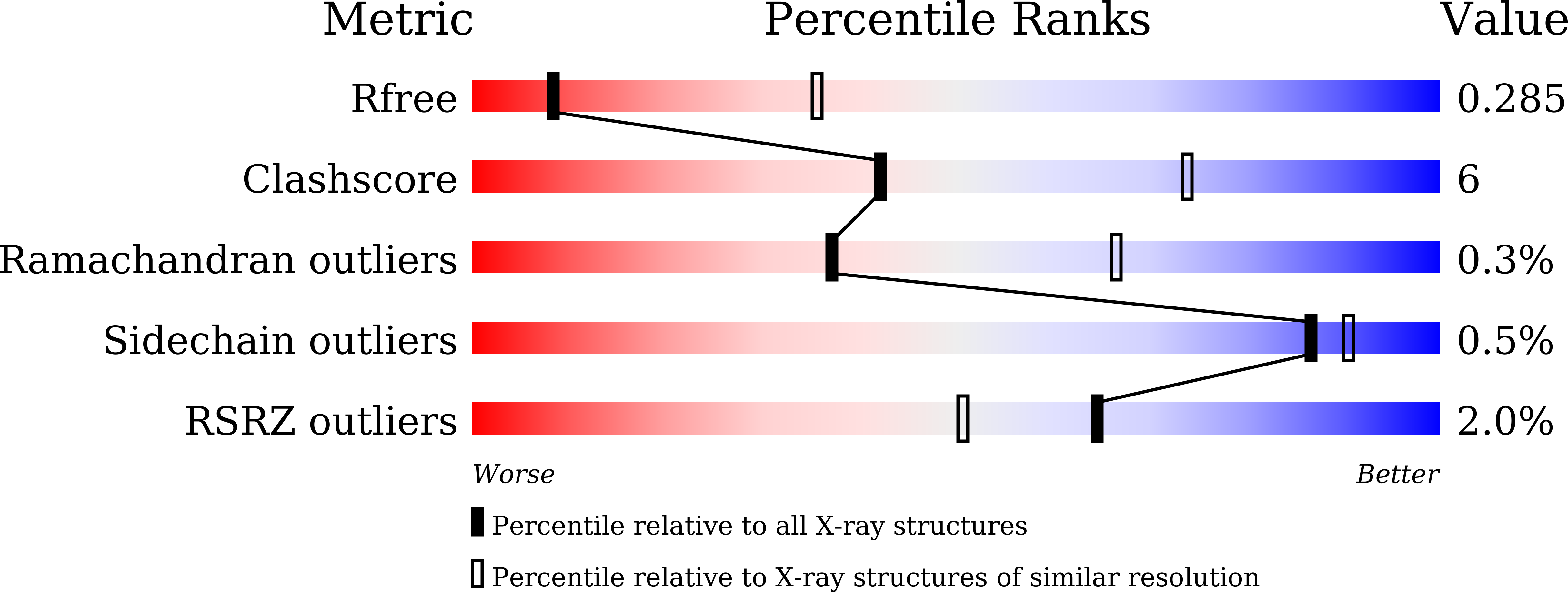
Resolution:
3.25 Å
R-Value Free:
0.28
R-Value Work:
0.24
R-Value Observed:
0.25
Space Group:
P 21 21 21