Deposition Date
2024-06-19
Release Date
2025-03-05
Last Version Date
2025-08-13
Entry Detail
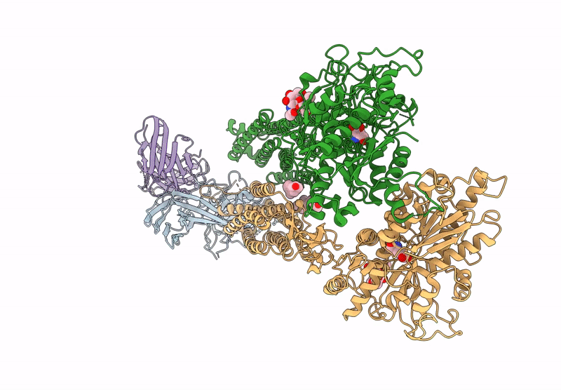
PDB ID:
9II3
Keywords:
Title:
Cryo-EM Structure of the 2:1 Complex of mGlu3 and beta-arrestin1
Biological Source:
Source Organism(s):
Homo sapiens (Taxon ID: 9606)
Mus musculus (Taxon ID: 10090)
Mus musculus (Taxon ID: 10090)
Expression System(s):
Method Details:
Experimental Method:
Resolution:
3.90 Å
Aggregation State:
PARTICLE
Reconstruction Method:
SINGLE PARTICLE