9IC7 image
Deposition Date 2025-02-14
Release Date 2025-12-03
Last Version Date 2025-12-03
Entry Detail
PDB ID:
9IC7
Keywords:
Title:
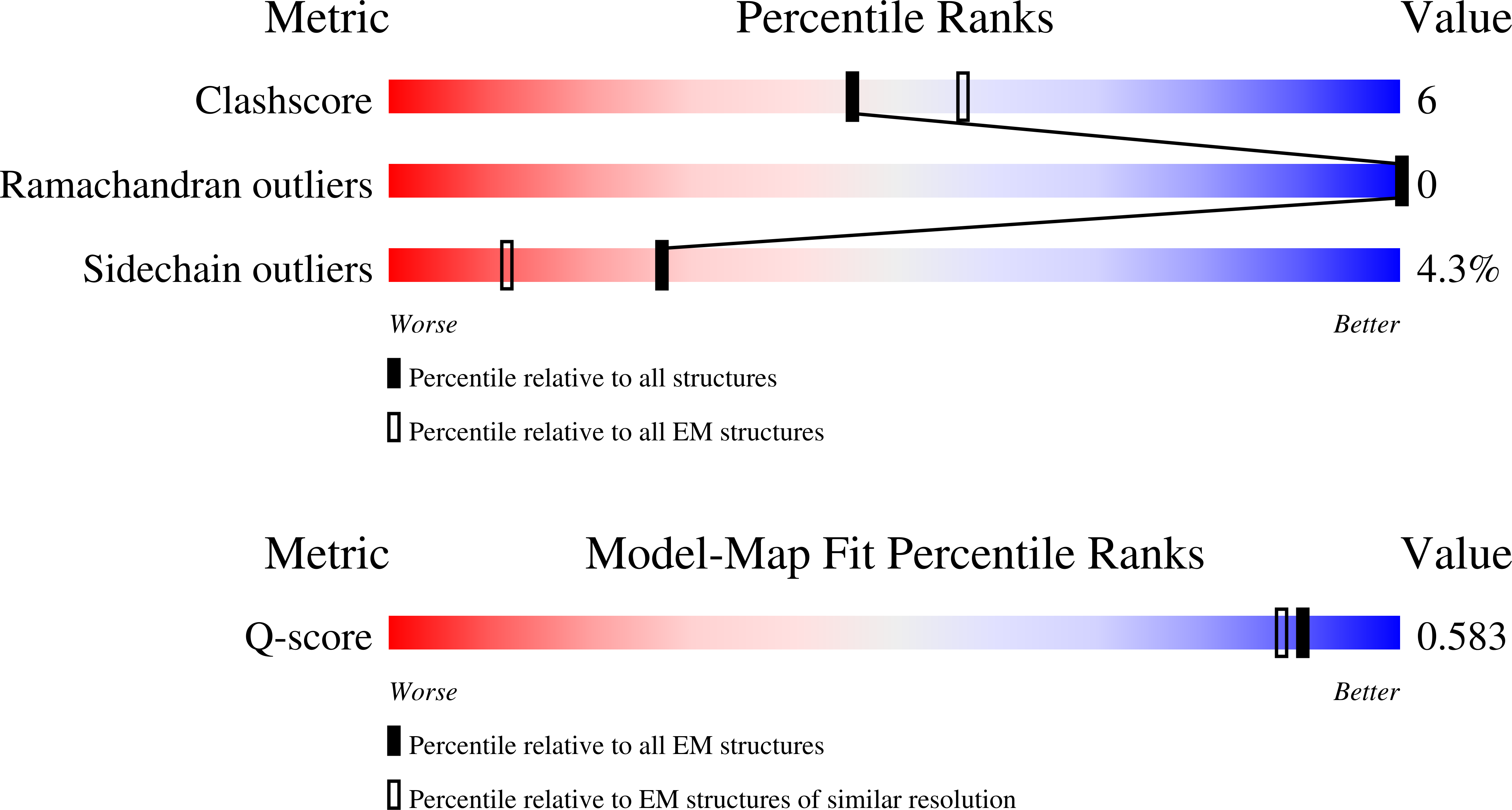
Cryo-EM structure of alpha-synuclein fibrils formed in artificial cerebrospinal fluid (aCSF)
Biological Source:
Source Organism:
Homo sapiens (Taxon ID: 9606)
Method Details:
Experimental Method:
Resolution:
2.90 Å
Aggregation State:
FILAMENT
Reconstruction Method:
HELICAL
Macromolecular Entities
Structures with similar UniProt ID
Protein Blast
Polymer Type:polypeptide(L)
Molecule:Alpha-synuclein
Gene (Uniprot):SNCA
Chain IDs:A (auth: n), B (auth: a), C (auth: b), D (auth: c), E (auth: d), F (auth: e), G (auth: f), H (auth: g), I (auth: h), J (auth: i), K (auth: j), L (auth: k), M (auth: l), N (auth: m)
Chain Length:140
Number of Molecules:14
Biological Source:Homo sapiens
Ligand Molecules
Primary Citation
Formation of Condition-Dependent Alpha-Synuclein Fibril Strain in Artificial Cerebrospinal Fluid.
Adv Sci ? e05228 e05228 (2025)
PMID: 41262012 DOI: 10.1002/advs.202505228

Abstact

α-Synuclein (aSyn) is an intrinsically disordered protein involved in neurotransmission and synaptic plasticity. The pathological aggregation of this protein is a hallmark of synucleinopathies such as Parkinson's disease (PD) or Multiple System Atrophy (MSA). Misfolded aSyn, which primarily originates in the cell cytosol, transmits between neurons, promoting a prion-like propagation. However, extracellular environments such as interstitial and cerebrospinal fluids (ISF & CSF) play a major role in its clearance and pathological transformation. The molecular components of CSF, including proteins, glycosaminoglycans, and metal ions, may influence the aggregate morphology, structure, and cytotoxicity to cells. To better understand how extracellular composition affects aggregates and their formation, artificial cerebrospinal fluid (aCSF) is employed to mimic potential aggregation processes occurring in CSF. Distinct aSyn fibrils are observed that exhibited low stability outside aCSF, and the removal of key CSF components led to its structural alterations. Cryo-electron microscopy revealed that these fibrils possess an electron density pocket coordinated with polar basic AAs (K43, K45, H50) that is also observed in aggregates obtained from PD and MSA patients. The findings illustrate the importance of physiologically relevant conditions in studying aSyn aggregation and may explain why disease-related fibril structure replication in vitro has not yet been successful.

Legend

Protein

Chemical

Disease

Primary Citation of related structures
Feedback Form
Name
Email
Institute
Feedback