9I4Z image
Deposition Date 2025-01-27
Release Date 2025-06-25
Last Version Date 2025-07-23
Entry Detail
PDB ID:
9I4Z
Keywords:
Title:
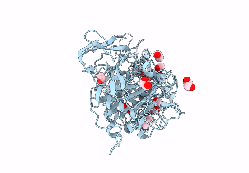
Crystal structure of Thomasclavelia ramosa IgA peptidase (IgAse) active site mutant (E330-N876)
Biological Source:
Source Organism:
Method Details:
Experimental Method:
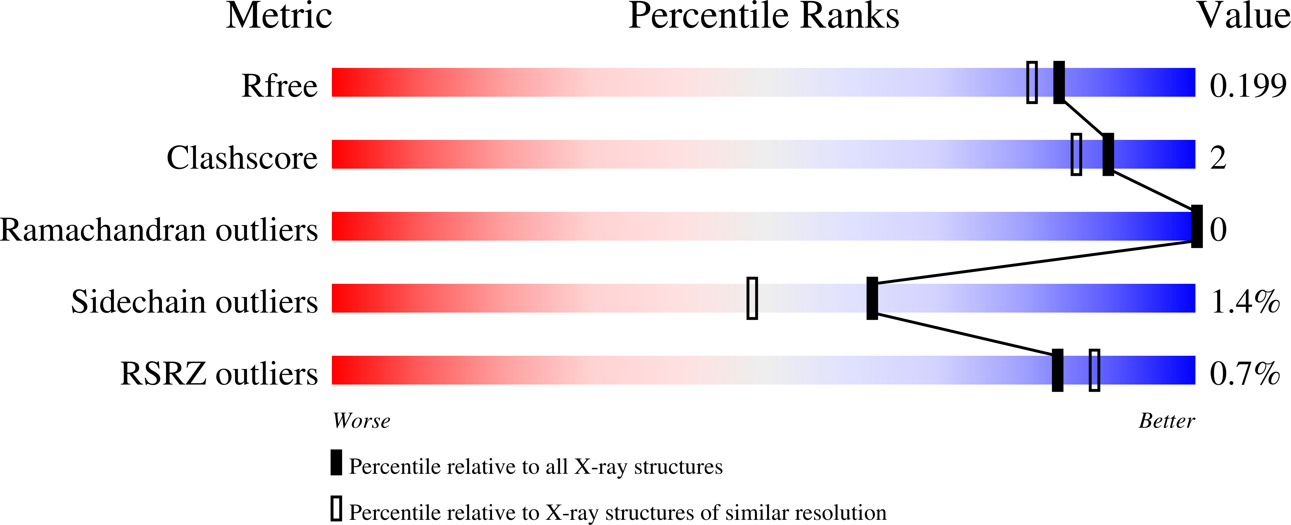
Resolution:
1.75 Å
R-Value Free:
0.21
R-Value Work:
0.17
R-Value Observed:
0.17
Space Group:
P 1 21 1
Macromolecular Entities
Structures with similar UniProt ID
Protein Blast
Polymer Type:polypeptide(L)
Molecule:IgA protease
Gene (Uniprot):iga
Mutagens:E540A
Chain IDs:A
Chain Length:559
Number of Molecules:1
Biological Source:Thomasclavelia ramosa
Primary Citation
Biochemical and structural characterization of the human gut microbiome metallopeptidase IgAse provides insight into its unique specificity for the Fab' region of IgA1 and IgA2.
Plos Pathog. 21 e1013292 e1013292 (2025)
PMID: 40627637 DOI: 10.1371/journal.ppat.1013292

Abstact

Human immunoglobulin A (IgA), comprising the isotypes IgA1 and IgA2, protects ~400 m2 of mucosal surfaces against microbial infections but can also lead to aberrant IgA deposits that cause disease. Certain bacteria have evolved peptidases that cleave the hinge between the Fab and Fc fragments of IgA, undermining its immune function. These peptidases specifically target IgA1, but not IgA2, which predominates in the gut and possesses a structurally distinct hinge region. The only known IgA2-specific peptidase is IgAse from the gut microbiome member Thomasclavelia ramosa, which also targets IgA1 but no other proteins. IgAse is a ~ 140-kDa, seven-domain, membrane-bound metallopeptidase (MP). Differential scanning fluorimetry, small-angle X-ray scattering, AI-based structural predictions, mass spectrometry, and high-resolution crystallography and cryo-electron microscopy of multidomain fragments of IgAse revealed a novel 313-residue catalytic domain (CD) from the igalysin family within the metzincin MP clan. The CD is flanked by an N-terminal globular C-type lectin-like domain and a wrapping domain (WD), followed by four all-β domains. Functional studies involving a comprehensive set of constructs (wild-type and mutant), authentic and recombinant IgA fragments, and inhibitors demonstrated that the minimal functional assembly requires the CD and WD, along with the Fab and hinge region (Fab'). Modelling studies suggested that the Fab heavy-chain constant domain interacts with the N-terminal subdomain of the CD, positioning the hinge peptide for cleavage-a mechanism confirmed by mutational analysis. These findings open avenues for therapeutic strategies to inhibit the only known IgA1/IgA2 peptidase and to develop it for dissolving pathologic IgA deposits.

Legend

Protein

Chemical

Disease

Primary Citation of related structures