9HPQ image
Deposition Date 2024-12-16
Release Date 2025-09-24
Last Version Date 2025-09-24
Entry Detail
PDB ID:
9HPQ
Keywords:
Title:
Peptide-substrate-binding (PSB) domain of human type I collagen prolyl 4-hydroxylase complexed with Pro-Pro-Gly-Pro-Arg-Gly-Pro-Pro-Gly.
Biological Source:
Source Organism:
Homo sapiens (Taxon ID: 9606)
Host Organism:
Method Details:
Experimental Method:
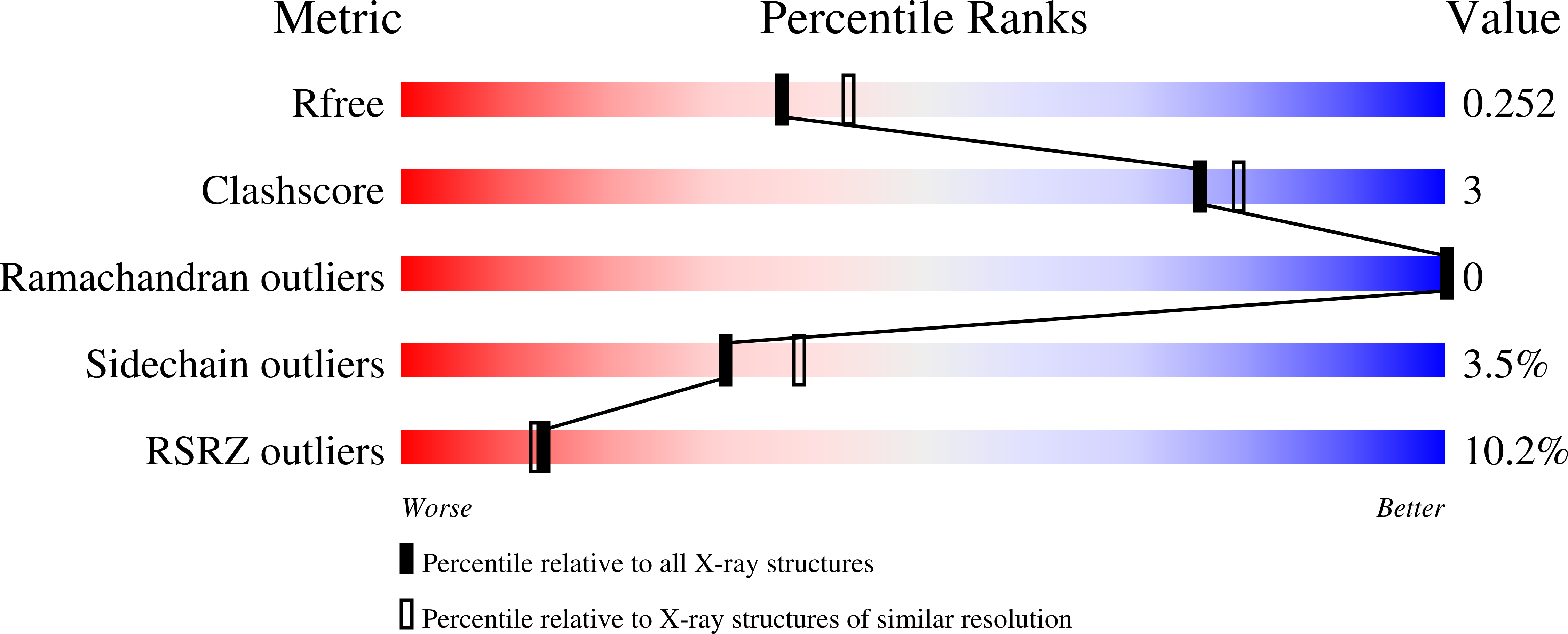
Resolution:
2.17 Å
R-Value Free:
0.24
R-Value Work:
0.22
Space Group:
P 21 21 2
Macromolecular Entities
Structures with similar UniProt ID
Protein Blast
Polymer Type:polypeptide(L)
Molecule:Prolyl 4-hydroxylase subunit alpha-1
Gene (Uniprot):P4HA1
Chain IDs:A, B, C, D
Chain Length:105
Number of Molecules:4
Biological Source:Homo sapiens
Polymer Type:polypeptide(L)
Molecule:Synthetic peptide PRO-PRO-GLY-PRO-ARG-GLY-PRO-PRO-GLY
Chain IDs:E, F, G
Chain Length:9
Number of Molecules:3
Biological Source:Homo sapiens
Primary Citation
Binding Differences of the Peptide-Substrate-Binding Domain of Collagen Prolyl 4-Hydroxylases I and II for Proline- and Hydroxyproline-Rich Peptides.
Proteins 93 1732 1746 (2025)
PMID: 40386874 DOI: 10.1002/prot.26839

Abstact

Collagen prolyl 4-hydroxylase (C-P4H) catalyzes the 4-hydroxylation of Y-prolines of the XYG-repeat of procollagen. C-P4Hs are tetrameric α2β2 enzymes. The α-subunit provides the N-terminal dimerization domain, the middle peptide-substrate-binding (PSB) domain, and the C-terminal catalytic (CAT) domain. There are three isoforms of the α-subunit, complexed with a β-subunit that is protein disulfide isomerase, forming C-P4H I-III. The PSB domain of the α-subunit binds proline-rich peptides, but its function with respect to the prolyl hydroxylation mechanism is unknown. An extended mode of binding of proline-rich peptides (PPII, polyproline type-II, conformation) to the PSB-I domain has previously been reported for the PPG-PPG-PPG and P9 peptides. Crystal structures now show that peptides with the motif PxGP (PPG-PRG-PPG, PPG-PAG-PPG) (where x, at Y-position 5, is not a proline) bind to the PSB-I domain differently, more deeply, in the peptide-binding groove. The latter mode of binding has previously been reported for structures of the PSB-II domain complexed with these PxGP-peptides. In addition, it is shown here by crystallographic binding studies that the POG-PAG-POG peptide (with 4-hydroxyprolines at Y-positions 2 and 8) also adopts the PxGP mode of binding to PSB-I as well as to PSB-II. Calorimetric binding studies show that the affinities of these peptides are lower for PSB-I than for PSB-II, with, respectively, KD values of about 70 μM for PSB-I and 20 μM for PSB-II. The importance of these results for understanding the reaction mechanism of C-P4H, in particular concerning the function of the PSB domain, is discussed.

Legend

Protein

Chemical

Disease

Primary Citation of related structures
Feedback Form
Name
Email
Institute
Feedback