9HNC image
Deposition Date 2024-12-10
Release Date 2025-05-14
Last Version Date 2025-05-14
Entry Detail
PDB ID:
9HNC
Keywords:
Title:
Crystal structure of potassium-independent L-asparaginase from Phaseolus vulgaris (PvAIII, PvAspG2)
Biological Source:
Source Organism:
Host Organism:
Method Details:
Experimental Method:
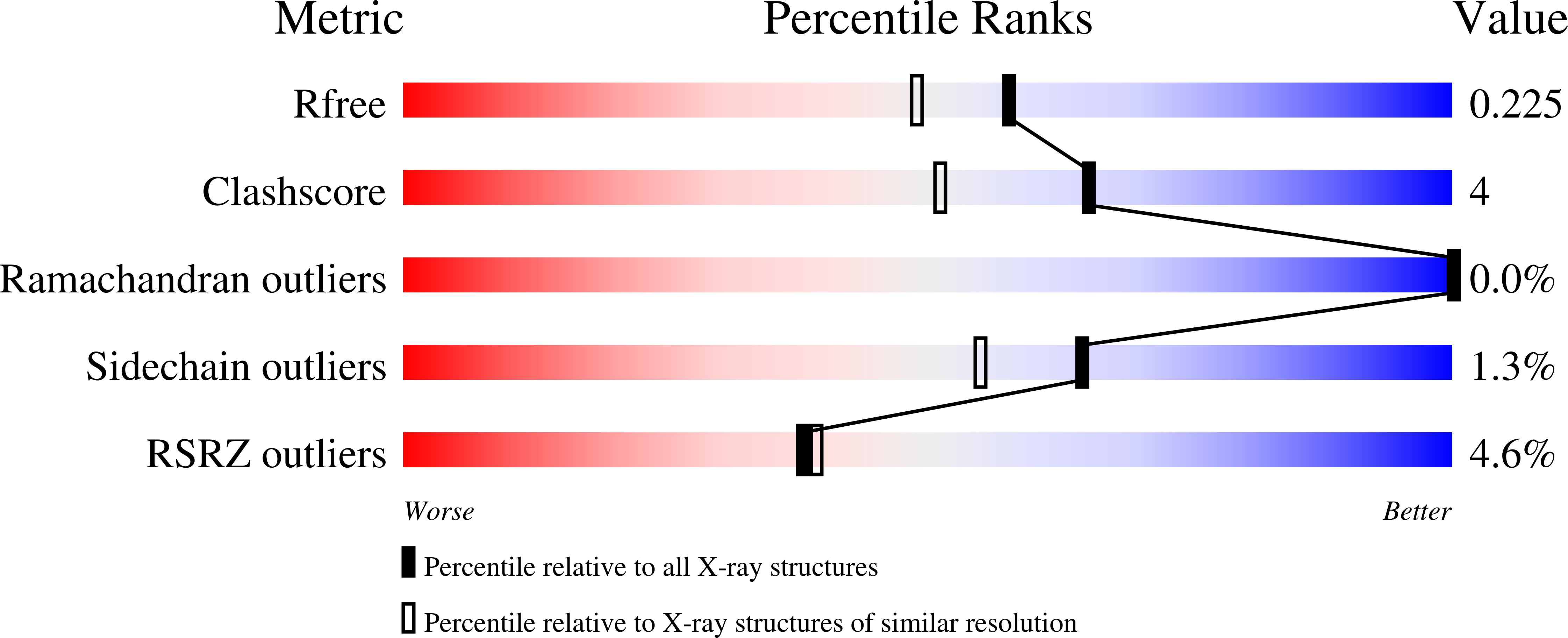
Resolution:
1.88 Å
R-Value Free:
0.21
R-Value Work:
0.18
Space Group:
P 1 2 1
Macromolecular Entities
Structures with similar UniProt ID
Protein Blast
Polymer Type:polypeptide(L)
Molecule:beta-aspartyl-peptidase
Gene (Uniprot):PHAVU_003G182400g
Chain IDs:A (auth: AAA), B (auth: BBB), C (auth: CCC), D (auth: DDD), E (auth: EEE), F (auth: FFF), G (auth: GGG), H (auth: HHH), I (auth: III), J (auth: JJJ), K (auth: KKK), L (auth: LLL), M (auth: MMM), N (auth: NNN), O (auth: OOO), P (auth: PPP)
Chain Length:322
Number of Molecules:16
Biological Source:Phaseolus vulgaris
Primary Citation
Unique double-helical packing of protein molecules in the crystal of potassium-independent L-asparaginase from common bean.
Acta Crystallogr D Struct Biol 81 252 264 (2025)
PMID: 40243630 DOI: 10.1107/S205979832500292X

Abstact

Common bean (Phaseolus vulgaris) encodes three class 2 L-asparaginase enzymes: two potassium-dependent enzymes [PvAIII(K)-1 and PvAIII(K)-2] and a potassium-independent enzyme (PvAIII). Here, we present the crystal structure of PvAIII, which displays a rare P2 space-group symmetry and a unique pseudosymmetric 41-like double-helical packing. The asymmetric unit contains 32 protein chains (16 αβ units labeled A-P) organized into two right-handed coiled arrangements, each consisting of four PvAIII (αβ)2 dimers. Detailed analysis of the crystal structure revealed that this unusual packing originates from three factors: (i) the ability of the PvAIII molecules to form extended intermolecular β-sheets, a feature enabled by the PvAIII sequence and secondary structure, (ii) incomplete degradation of the flexible linker remaining at the C-terminus of α subunits of protein chain C after the autoproteolytic cleavage (maturation) of the PvAIII precursor and (iii) intermolecular entanglement between protein chains from the two helices to create `hydrogen-bond linchpins' that connect adjacent protein chains. The Km value of PvAIII for L-asparagine is approximately five times higher than for β-peptides, suggesting that the physiological role of PvAIII may be more related to the removal of toxic β-peptides than to basic L-asparagine metabolism. A comparison of the active sites of PvAIII and PvAIII(K)-1 shows that the proteins have nearly identical residues in the catalytic center, except for Thr219, which is unique to PvAIII. To test whether the residue type at position 219 affects the enzymatic activity of PvAIII, we designed and produced a T219S mutant. The kinetic parameters determined for L-asparagine hydrolysis indicate that the T/S residue type at position 219 does not affect the L-asparaginase activity of PvAIII.

Legend

Protein

Chemical

Disease

Primary Citation of related structures