9HMX image
Deposition Date 2024-12-09
Release Date 2025-07-30
Last Version Date 2025-07-30
Entry Detail
PDB ID:
9HMX
Keywords:
Title:
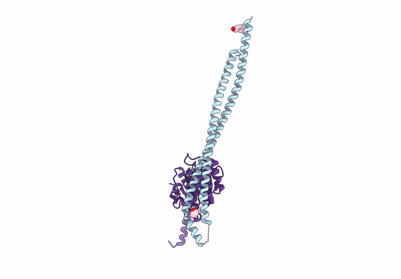
Structure of SteB-RipA complex from Mycobacterium tuberculosis
Biological Source:
Source Organism:
Method Details:
Experimental Method:
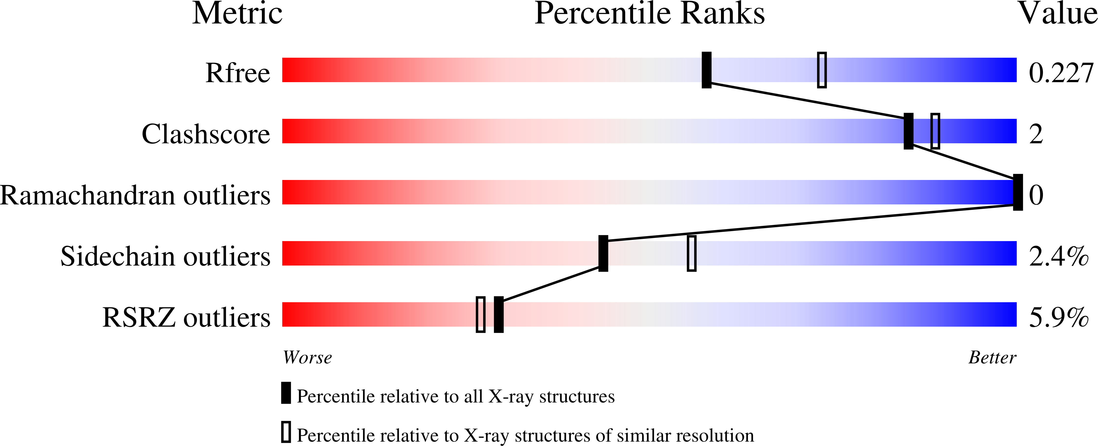
Resolution:
2.22 Å
R-Value Free:
0.23
R-Value Work:
0.20
R-Value Observed:
0.20
Space Group:
C 1 2 1
Macromolecular Entities
Structures with similar UniProt ID
Protein Blast
Polymer Type:polypeptide(L)
Molecule:Peptidoglycan endopeptidase RipA
Gene (Uniprot):ripA
Chain IDs:A
Chain Length:216
Number of Molecules:1
Biological Source:Mycobacterium tuberculosis H37Rv
Structures with similar UniProt ID
Protein Blast
Polymer Type:polypeptide(L)
Molecule:Copper transporter MctB
Gene (Uniprot):mctB
Chain IDs:B
Chain Length:277
Number of Molecules:1
Biological Source:Mycobacterium tuberculosis H37Rv
Ligand Molecules
Primary Citation
Mechanistic insights into the allosteric regulation of cell wall hydrolase RipA in Mycobacterium tuberculosis.
Biorxiv ? ? ? (2025)
PMID: 40667032 DOI: 10.1101/2025.06.28.662095

Abstact

D,L-endopeptidase RipA is the major PG hydrolase required for daughter cell separation in Mycobacterium tuberculosis (Mtb), as RipA defects severely hinder cell division and increase antibiotic vulnerability. Despite extensive studies, the mechanisms governing Mtb RipA regulation remain controversial and poorly understood. Here, we report an integrative structural and functional analysis of the SteAB system, a regulatory complex that has been shown to modulate cell separation in the model organism Corynebacterium glutamicum (Cglu) and is conserved across Mycobacteriales . Although Mtb SteB was previously proposed to act as a mycobacterial outer membrane copper transporter, the crystal structures of the homodimeric protein, alone and in complex with the RipA coiled-coil (CC) domain, rule out this hypothesis. Instead, the high-affinity SteB-RipA interaction, together with computational and biophysical data, strongly supports the role of SteB as a direct RipA activator that releases enzyme autoinhibition upon complex formation. In addition, crystallographic characterization of the cytoplasmic core of SteA revealed a homodimeric organization harboring a conserved functional pocket similar to the phosphonucleotide-binding site of thiamine pyrophosphokinase. These data, coupled with the in vivo phenotypic analysis of a steAB knockout mutant of Cglu , support a model in which the transmembrane SteAB heterotetramer, driven by cytoplasmic ligand binding, orchestrates the productive periplasmic positioning of RipA, leading to PG hydrolysis activation. These findings shed new light on the regulation of mycobacterial cell wall remodeling, with implications for understanding Mtb pathogenesis and identifying novel antimicrobial targets.

Legend

Protein

Chemical

Disease

Primary Citation of related structures
Feedback Form
Name
Email
Institute
Feedback