9HM2 image
Deposition Date 2024-12-06
Release Date 2025-10-22
Last Version Date 2026-01-28
Entry Detail
PDB ID:
9HM2
Keywords:
Title:
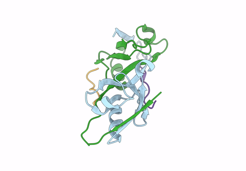
A swapped dimeric form of ZO1/TJP1 PDZ2 in complex with the C-terminal peptide from protein E of SARS-CoV-2
Biological Source:
Expression System(s):
Method Details:
Experimental Method:
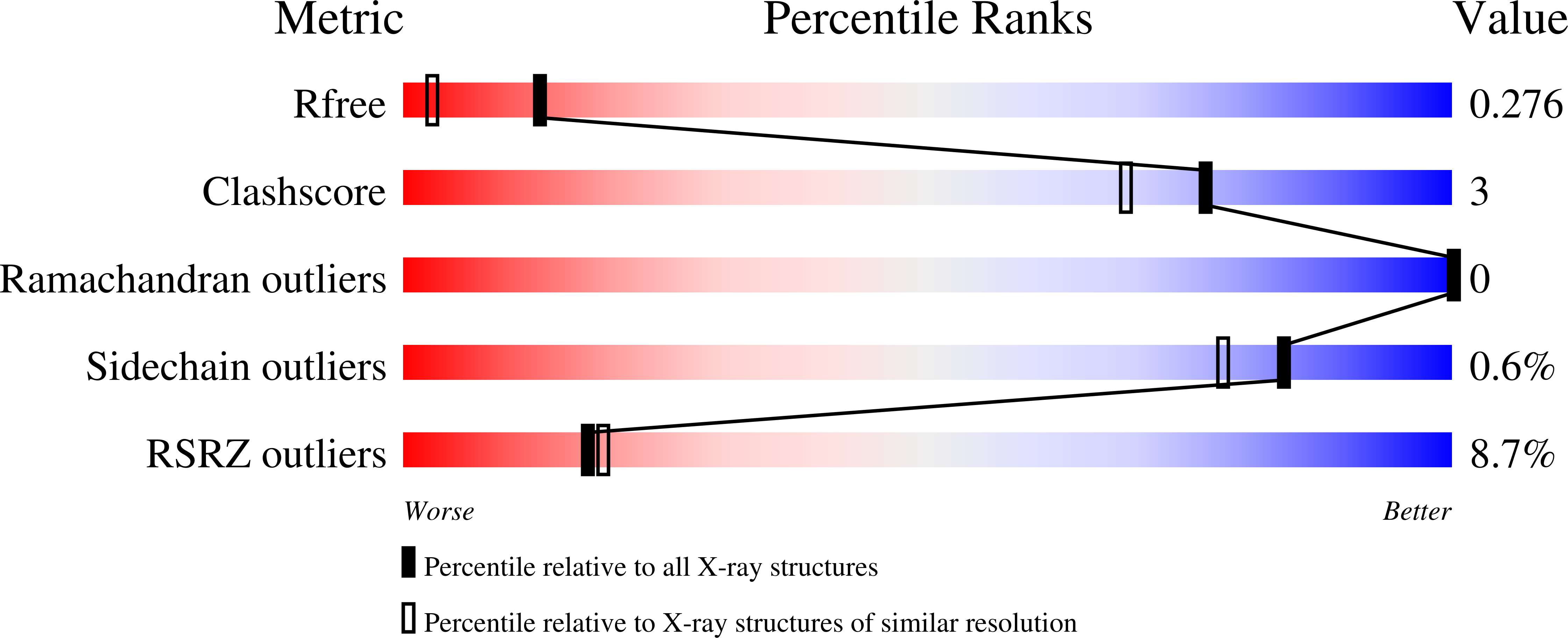
Resolution:
1.72 Å
R-Value Free:
0.27
R-Value Work:
0.20
R-Value Observed:
0.20
Space Group:
P 21 21 21
Macromolecular Entities
Structures with similar UniProt ID
Protein Blast
Polymer Type:polypeptide(L)
Molecule:Tight junction protein ZO-1
Gene (Uniprot):TJP1
Chain IDs:A, B
Chain Length:84
Number of Molecules:2
Biological Source:Homo sapiens
Structures with similar UniProt ID
Protein Blast
Polymer Type:polypeptide(L)
Molecule:Envelope small membrane protein
Gene (Uniprot):E
Chain IDs:C, D
Chain Length:12
Number of Molecules:2
Biological Source:Severe acute respiratory syndrome coronavirus 2
Ligand Molecules
Primary Citation

Abstact

The SARS-CoV-2 E protein through its C-terminal PDZ-binding motif (PBM) interacts with several host PDZ-containing proteins, including Zonula occludens-1 (ZO-1) protein via its PDZ2 domain, thereby contributing to viral pathogenesis. Targeting this interaction represents a potential therapeutic strategy. In this study, we determined the X-ray structure of the E PBM peptide in complex with the ZO-1 PDZ2 domain at 1.7 Å resolution. The structure revealed a domain-swapped dimer conformation of ZO-1 PDZ2, with the E PBM peptide conventionally bound within the PDZ domain's canonical binding groove, exhibiting key interactions characteristic of type II PBM/PDZ interactions. To identify potential inhibitors of the E PBM/ZO-1 PDZ2 interaction, we performed a Homogeneous Time-Resolved Fluorescence (HTRF) screening using a protein-protein interaction-focused library of 1000 compounds. This led to the identification of 36 hits that disrupted this interaction. Subsequent cytotoxicity and dose-response assays narrowed the selection to 14 promising compounds. Docking simulations showed that some compounds bind within or near the PBM-binding pocket, supporting a competitive mechanism of interaction inhibition, while others bind at a central interface between the two PDZ monomers, suggesting an inhibition of dimerization, which in turn prevents PBM binding. Thus, the E PBM/ZO-1 PDZ2 interaction can be inhibited through both direct and indirect mechanisms. Finally, antiviral assays using a NanoLuciferase-expressing recombinant SARS-CoV-2 demonstrated that one compound, C19, significantly reduced viral replication, highlighting its potential as a candidate for further therapeutic development.

Legend

Protein

Chemical

Disease

Primary Citation of related structures
Feedback Form
Name
Email
Institute
Feedback