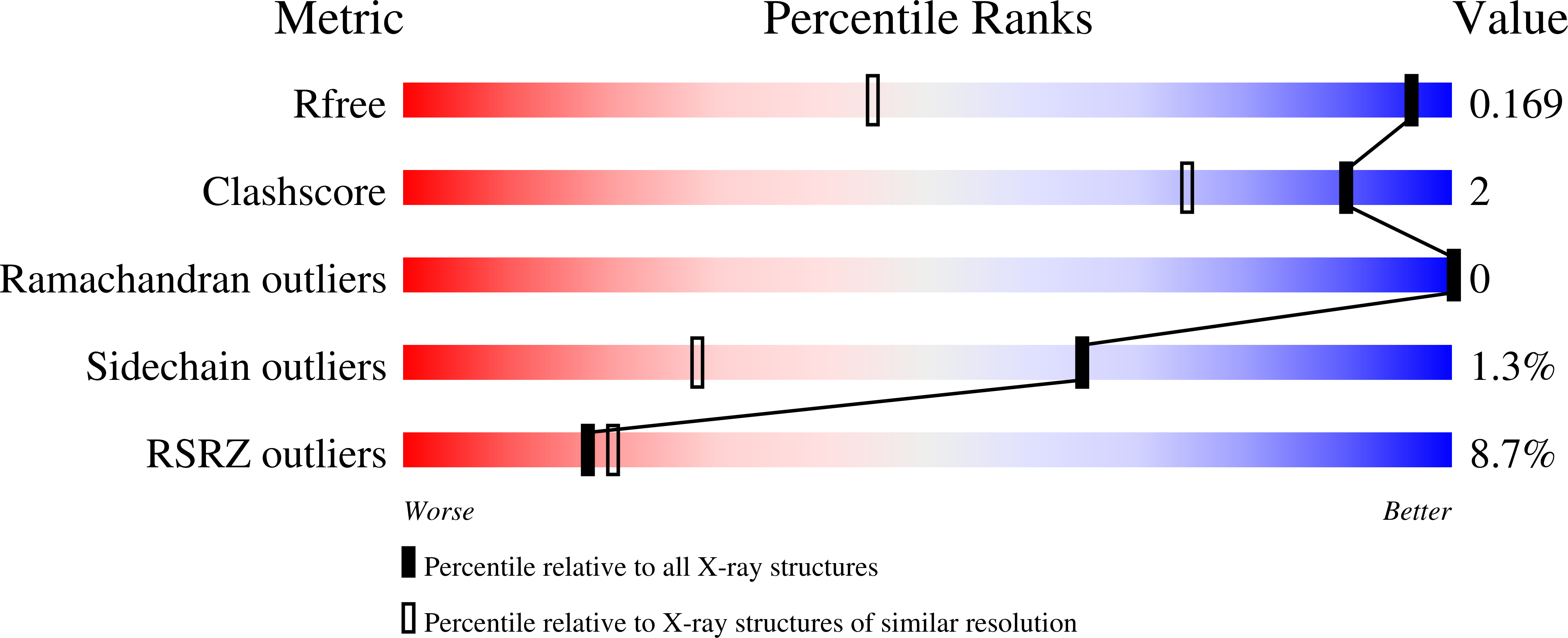
9H48 image
Deposition Date 2024-10-17
Release Date 2024-12-11
Last Version Date 2024-12-11
Entry Detail
PDB ID:
9H48
Keywords:
Title:
Mouse Iodothyronine deiodinase 2 catalytic core, mutant - LysLys180AlaAla, Secys-> Cys
Biological Source:
Source Organism:
Mus musculus (Taxon ID: 10090)
Host Organism:
Method Details:
Experimental Method:
Resolution:
1.09 Å
R-Value Free:
0.16
R-Value Work:
0.14
R-Value Observed:
0.14
Space Group:
P 32
Macromolecular Entities
Structures with similar UniProt ID
Protein Blast
Polymer Type:polypeptide(L)
Molecule:Type II iodothyronine deiodinase
Gene (Uniprot):Dio2
Mutagens:K180A, K181A, U126C
Chain IDs:A
Chain Length:193
Number of Molecules:1
Biological Source:Mus musculus
Primary Citation
Structural Insights into the Iodothyronine Deiodinase 2 Catalytic Core and Deiodinase Catalysis and Dimerization.
Biomolecules 14 ? ? (2024)
PMID: 39595550 DOI: 10.3390/biom14111373

Abstact

Iodothyronine deiodinases (Dio) are selenocysteine-containing membrane enzymes that activate and inactivate the thyroid hormones (TH) through reductive iodide eliminations. The three deiodinase isoforms are homodimers sharing highly conserved amino acid sequences, but they differ in their regioselectivities for the deiodination reaction and regulatory features. We have now solved a crystal structure of the mouse deiodinase 2 (Dio2) catalytic domain. It reveals a high overall similarity to the deiodinase 3 structure, supporting the proposed common mechanism, but also Dio2-specific features, likely mediating its unique properties. Activity studies with an artificially enforced Dio dimer further confirm that dimerization is required for activity and requires both the catalytic core and the enzyme's N-terminus. Cross-linking studies reveal the catalytic core's dimerization interface, providing insights into the architecture of the complete, active Dio homodimer.

Legend

Protein

Chemical

Disease

Primary Citation of related structures