9H1M image
Entry Detail
PDB ID:
9H1M
Keywords:
Title:
Recombinant ferric horseradish peroxidase C1A
Biological Source:
Source Organism:
PDB Version:
Deposition Date:
2024-10-09
Release Date:
2025-03-19
Method Details:
Experimental Method:
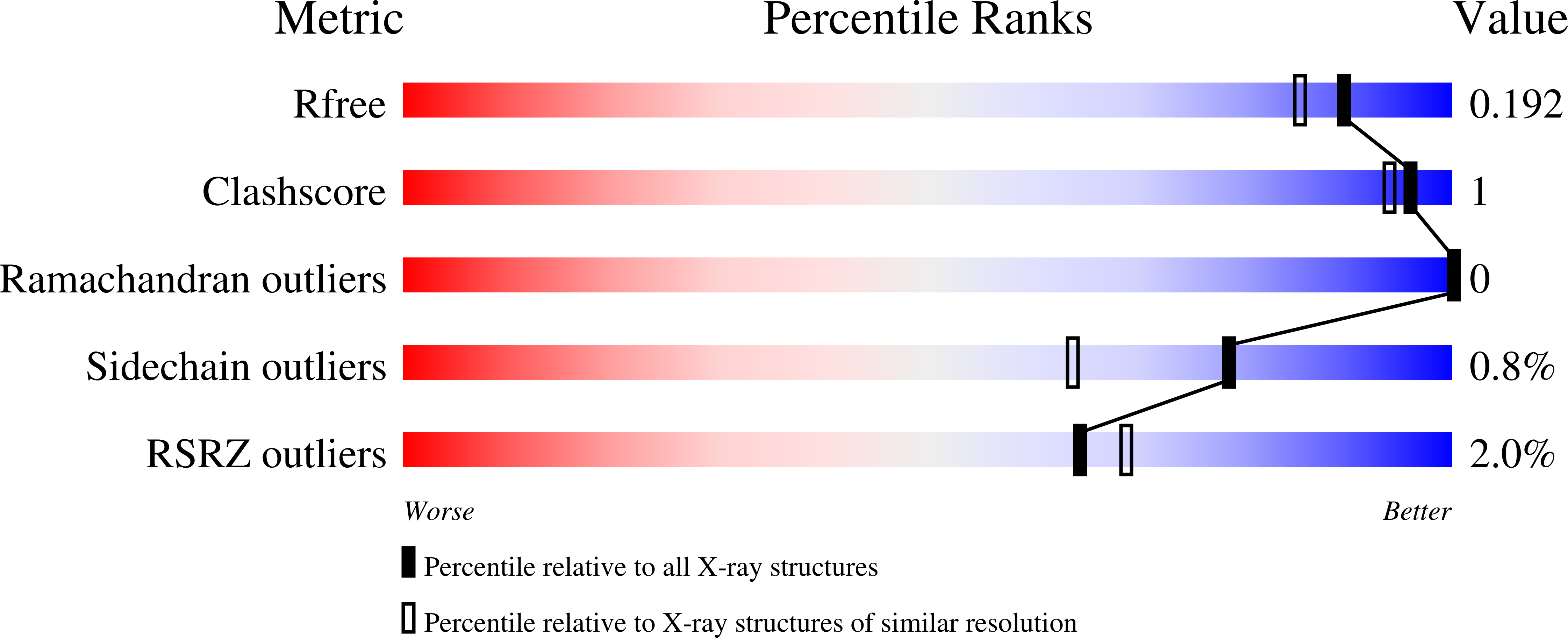
Resolution:
1.63 Å
R-Value Free:
0.18
R-Value Work:
0.16
R-Value Observed:
0.17
Space Group:
P 21 21 21
Macromolecular Entities
Polymer Type:polypeptide(L)
Description:Peroxidase C1A
Chain IDs:A
Chain Length:309
Number of Molecules:1
Biological Source:Armoracia rusticana
Primary Citation
Crystal structure of ferric recombinant horseradish peroxidase.
J.Biol.Inorg.Chem. 30 221 227 (2025)
PMID: 40053124 DOI: 10.1007/s00775-025-02103-2

Abstact

Horseradish peroxidase (HRP), isolated from horseradish roots, is heavily glycosylated, making it difficult to crystallize. In this work, we produced recombinant HRP in E. coli and obtained an X-ray structure of the ferric enzyme at 1.63 Å resolution. The structure shows that the recombinant HRP contains four disulphide bonds and two calcium ions, which are highly conserved in class III peroxidase enzymes. The heme active site contains histidine residues at the proximal (His 170) and distal (His 42) positions, and an active site arginine (Arg 38). Surprisingly, an ethylene glycol molecule was identified in the active site, forming hydrogen bonds with His 42 and Arg 38 at the δ-heme edge. The high yields obtained from the recombinant expression system, and the successful crystallization of the enzyme pave the way for new structural studies in the future.

Legend

Protein

Chemical

Disease

Primary Citation of related structures