9GV7 image
Entry Detail
PDB ID:
9GV7
Keywords:
Title:
Structure of reverse docking TCR in complex with peptide-HLA
Biological Source:
Source Organism:
Host Organism:
PDB Version:
Deposition Date:
2024-09-23
Release Date:
2025-04-23
Method Details:
Experimental Method:
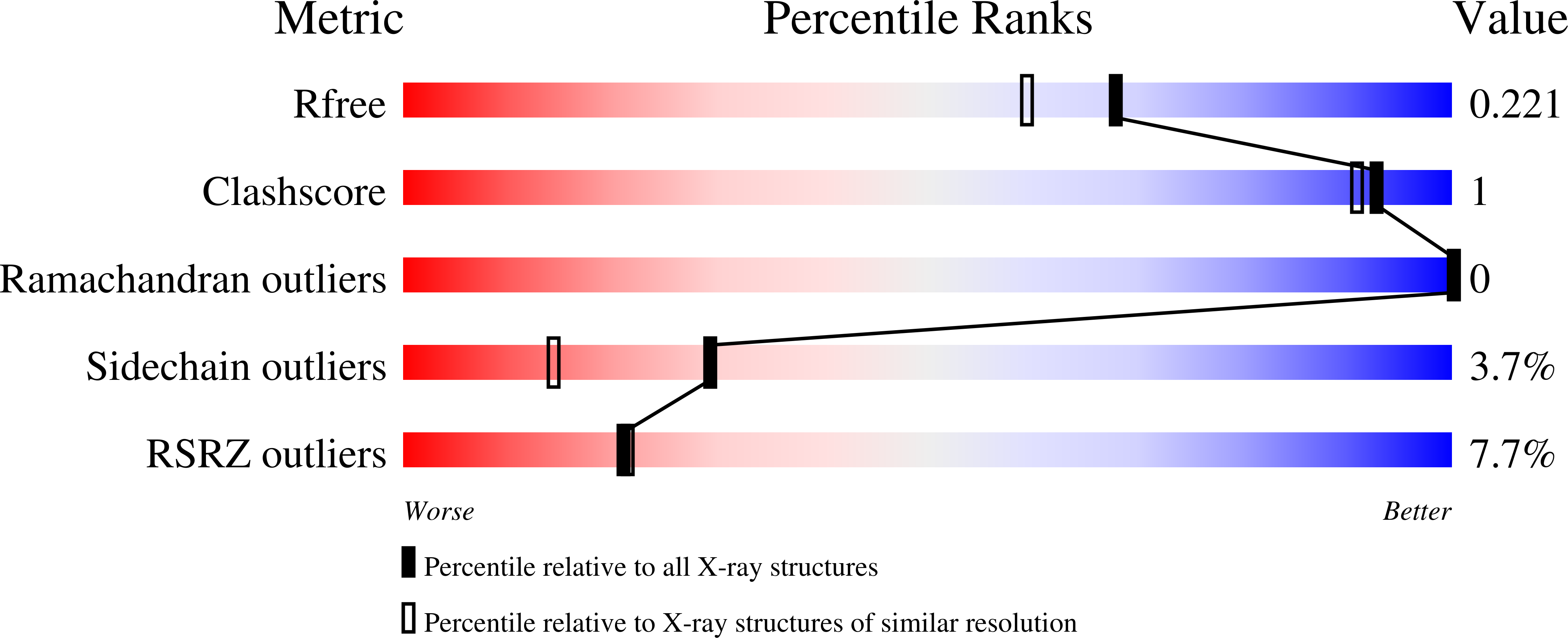
Resolution:
1.86 Å
R-Value Free:
0.21
R-Value Work:
0.18
R-Value Observed:
0.18
Space Group:
P 65
Macromolecular Entities
Polymer Type:polypeptide(L)
Description:MHC class I antigen
Chain IDs:A
Chain Length:276
Number of Molecules:1
Biological Source:Homo sapiens
Polymer Type:polypeptide(L)
Description:Beta-2-microglobulin
Chain IDs:B
Chain Length:100
Number of Molecules:1
Biological Source:Homo sapiens
Polymer Type:polypeptide(L)
Description:Peptide
Chain IDs:C
Chain Length:11
Number of Molecules:1
Biological Source:Homo sapiens
Polymer Type:polypeptide(L)
Description:TCR Alpha
Chain IDs:D
Chain Length:200
Number of Molecules:1
Biological Source:Homo sapiens
Polymer Type:polypeptide(L)
Description:TCR Beta
Chain IDs:E
Chain Length:244
Number of Molecules:1
Biological Source:Homo sapiens
Ligand Molecules
Primary Citation
Determining T-cell receptor binding orientation and Peptide-HLA interactions using cross-linking mass spectrometry.
J.Biol.Chem. 301 108445 108445 (2025)
PMID: 40154610 DOI: 10.1016/j.jbc.2025.108445

Abstact

T cell receptors (TCRs) recognize specific peptides presented by human leukocyte antigens (HLAs) on the surface of antigen-presenting cells and are involved in fighting pathogens and cancer surveillance. Canonical docking orientation of TCRs to their target peptide-HLAs (pHLAs) is essential for T cell activation, with reverse binding TCRs lacking functionality. TCR binding geometry and molecular interaction footprint with pHLAs are typically obtained by determining the crystal structure. Here, we describe the use of a cross-linking tandem mass spectrometry (XL-MS/MS) method to decipher the binding orientation of several TCRs to their target pHLAs. Cross-linking sites were localized to specific residues and their molecular interactions showed differentiation between TCRs binding in canonical or reverse orientations. Structural prediction and crystal structure determination of two TCR-pHLA complexes validated these findings. The XL-MS/MS method described herein offers a faster and simpler approach for elucidating TCR-pHLA binding orientation and interactions.

Legend

Protein

Chemical

Disease

Primary Citation of related structures