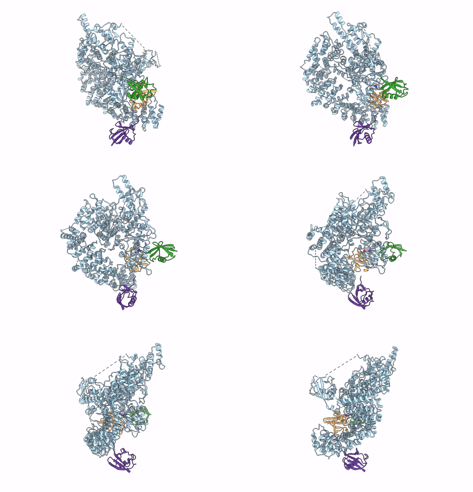
9GKM image
Deposition Date 2024-08-25
Release Date 2025-05-21
Last Version Date 2025-06-04
Entry Detail
PDB ID:
9GKM
Keywords:
Title:
Structure of HECT E3 TRIP12 forming K29/K48-branched Ubiquitin chains
Biological Source:
Source Organism:
Homo sapiens (Taxon ID: 9606)
Host Organism:
Method Details:
Experimental Method:
Resolution:
3.69 Å
Aggregation State:
PARTICLE
Reconstruction Method:
SINGLE PARTICLE
Macromolecular Entities
Polymer Type:polypeptide(L)
Molecule:Isoform 3 of E3 ubiquitin-protein ligase TRIP12
Gene (Uniprot):TRIP12
Chain IDs:D (auth: A)
Chain Length:1565
Number of Molecules:1
Biological Source:Homo sapiens
Polymer Type:polypeptide(L)
Molecule:Ubiquitin
Gene (Uniprot):UBC
Chain IDs:A (auth: B)
Chain Length:75
Number of Molecules:1
Biological Source:Homo sapiens
Polymer Type:polypeptide(L)
Molecule:Polyubiquitin-C
Gene (Uniprot):UBC
Mutations:K29C
Chain IDs:B (auth: C)
Chain Length:76
Number of Molecules:1
Biological Source:Homo sapiens
Polymer Type:polypeptide(L)
Molecule:Polyubiquitin-B
Mutations:K48R
Chain IDs:C (auth: D)
Chain Length:76
Number of Molecules:1
Biological Source:Homo sapiens
Ligand Molecules
Primary Citation
TRIP12 structures reveal HECT E3 formation of K29 linkages and branched ubiquitin chains.
Nat.Struct.Mol.Biol. ? ? ? (2025)
PMID: 40419785 DOI: 10.1038/s41594-025-01561-1

Abstact

Regulation by ubiquitin depends on E3 ligases forging chains of specific topologies, yet the mechanisms underlying the generation of atypical linkages remain largely elusive. Here we utilize biochemistry, chemistry, and cryo-EM to define the catalytic architecture producing K29 linkages and K29/K48 branches for the human HECT E3 TRIP12. TRIP12 resembles a pincer. One pincer side comprises tandem ubiquitin-binding domains, engaging the proximal ubiquitin to direct its K29 towards the ubiquitylation active site, and selectively capturing a distal ubiquitin from a K48-linked chain. The opposite pincer side-the HECT domain-precisely juxtaposes the ubiquitins to be joined, further ensuring K29 linkage specificity. Comparison to the prior structure visualizing K48-linked chain formation by UBR5 reveals a similar mechanism shared by two human HECT enzymes: parallel features of the E3s, donor and acceptor ubiquitins configure the active site around the targeted lysine, with E3-specific domains buttressing the acceptor for linkage-specific polyubiquitylation.

Legend

Protein

Chemical

Disease

Primary Citation of related structures